Treatment of subarachnoid hemorrhage in the brain. Types of subarachnoid hemorrhage Subarachnoid cerebral hemorrhage in a child
Subarachnoid hemorrhage is a type of intracranial hemorrhage in which blood spreads into the subarachnoid space of the brain and spinal cord. A distinction is made between subarachnoid hemorrhage in TBI and in acute cerebrovascular accident of the hemorrhagic type. To refer to the latter, the terms “spontaneous subarachnoid hemorrhage” and “non-traumatic subarachnoid hemorrhage” are used.
ICD-I0 codes: 160.0-160.9. Subarachnoid hemorrhage.
EPIDEMIOLOGY
According to stroke registries in different countries, the incidence of subarachnoid hemorrhage is 14-20 per 100,000 population per year. The proportion of subarachnoid hemorrhage among other types of stroke does not exceed 5%.
Subarachnoid hemorrhage can happen at any age, but it most often occurs between 40 and 60 years of age.
ETIOLOGY
The causes of subarachnoid hemorrhage are varied, but most often it is a consequence of rupture of aneurysms of cerebral vessels, accounting for 70-80% of all subarachnoid hemorrhages. Diseases in which subarachnoid hemorrhage may develop are listed below.
Primary vascular diseases of the central nervous system:
- arterial aneurysms of cerebral vessels;
- vascular malformations of the central nervous system (arteriovenous malformations, cavernomas, arteriovenous fistulas);
- anomalies of the vascular system of the brain (Nishimoto disease, dissecting aneurysms of cerebral vessels).
Secondary vascular pathology of the central nervous system:
- arterial hypertension;
- vasculitis;
- blood diseases;
- violation of the blood coagulation system when taking anticoagulants, antiplatelet agents, contraceptives and other medications.
When it is not possible to establish the etiological factor of subarachnoid hemorrhage, the concept of “subarachnoid hemorrhage of unknown origin” is used. The share of such hemorrhages accounts for about 15%.
CLASSIFICATION
Subarachnoid hemorrhages are classified according to etiological factor and prevalence. The latter is possible only on the basis of CT or MRI data. In this case, both the massiveness of the hemorrhage and its combination with other components of intracranial hemorrhage - parenchymal and ventricular - are taken into account. Depending on this factor, isolated subarachnoid hemorrhage, subarachnoid-parenchymal, subarachnoid-ventricular and subarachnoid-parenchymal-ventricular hemorrhage are distinguished (Fig. 30-6).
Rice. 30- 6. Typical subarachnoid hemorrhage. A symmetrical distribution of blood is visible in the basal cisterns, interhemispheric fissure, and convexital subarachnoid spaces (CT).
In world practice, the classification of subarachnoid hemorrhages proposed by M. Fisher (1980) has become widespread. It characterizes the prevalence of subarachnoid hemorrhage according to CT results (Table 30-1)
Table 30-1. Classification of hemorrhage according to M. Fisher (1980)
CLINICAL PICTURE
Subarachnoid hemorrhage develops acutely, without any precursors, and is characterized by the occurrence of a sudden intense diffuse headache of the “blow” type, “spreading of hot liquid in the head.” nausea, vomiting. Short-term loss of consciousness and rapid development of meningeal syndrome in the absence of focal neurological disorders are typical.
Prolonged loss of consciousness indicates severe hemorrhage, usually with a breakthrough of blood into the ventricular system, and the rapid onset of focal symptoms indicates subarachnoid-parenchymal hemorrhage.
Meningeal symptoms are the main differential diagnostic sign of subarachnoid hemorrhage. Depending on the massiveness of the subarachnoid hemorrhage, they can be expressed to varying degrees and persist from several days to 3-4 weeks.
Along with the development of neurological symptoms, subarachnoid hemorrhage can be accompanied by various viscerovegetative disorders. Most often, at the time of hemorrhage, an increase in blood pressure is recorded. Increased blood pressure is a reaction to a stressful situation. at the same time having a compensatory nature, since it ensures the maintenance of cerebral perfusion pressure in conditions of intracranial hypertension that occurs at the time of subarachnoid hemorrhage. High blood pressure at the time of hemorrhage, especially in patients suffering from arterial hypertension, can cause an erroneous interpretation of the acute condition as a hypertensive crisis.
In cases of severe subarachnoid hemorrhage, cardiac and respiratory disturbances may occur.
In the acute stage of subarachnoid hemorrhage, an increase in body temperature up to febrile levels, as well as the development of leukocytosis, are often noted.
These symptoms may be misinterpreted as signs of an infectious disease.
The severity of the patient's condition at the time of subarachnoid hemorrhage and the further course of the disease depend primarily on the massiveness of the hemorrhage and its etiology. Subarachnoid hemorrhages occur most severely when cerebral aneurysms rupture (see section “Surgical treatment of cerebral aneurysms”).
DIAGNOSTICS
The clinical diagnosis of subarachnoid hemorrhage must be confirmed by instrumental studies. The most reliable and accessible method for diagnosing subarachnoid hemorrhage to date remains lumbar puncture. The cerebrospinal fluid in subarachnoid hemorrhage is intensely stained with blood. An admixture of blood in the cerebrospinal fluid, gradually decreasing. persists for 1-2 weeks from the onset of the disease. Subsequently, the cerebrospinal fluid acquires a xanthochromic color.
In unconscious patients, lumbar puncture should be performed with great caution due to the risk of brain dislocation.
In recent years, CT has become the method of choice in the diagnosis of subarachnoid hemorrhage. CT not only detects and evaluates the prevalence of blood in the subarachnoid space, but also allows one to obtain information about the presence of ventricular and parenchymal components of hemorrhage, edema and dislocation of the brain, and the state of the cerebrospinal fluid system. Without these data, correct management of a patient with subarachnoid hemorrhage at the present stage of development of neurosurgery is impossible. In some cases, even with a conventional CT scan it is possible to establish or suggest the cause of hemorrhage. Modern computed tomographs also make it possible to perform high-quality examination of the cerebral vascular system (CT angiography), which provides more than 90% accuracy in diagnosing the source of bleeding.
When CT diagnostics of subarachnoid hemorrhage, it is necessary to take into account that the information content of the method is directly dependent on the timing of the CT scan (time elapsed after the hemorrhage), which is due to changes in the radiopaque properties of the shed blood. Already a week after subarachnoid hemorrhage, blood in the subarachnoid space is visible only in half of the cases. In this regard, with negative CT data, patients with a clinical picture of subarachnoid hemorrhage require a diagnostic lumbar puncture.
Diagnosis of subarachnoid hemorrhage using MRI is less accurate due to rapid changes in signal intensity caused by the transformation of hemoglobin molecules in the erupted blood. However, in the absence of CT, MRI can be successfully used not only to diagnose subarachnoid hemorrhage, but also to determine the source of bleeding (MRI angiography). To diagnose vasospasm, one of the complications of subarachnoid hemorrhage, TCD is used. This study allows us to identify vasospasm in the vessels of the base of the brain, determine its prevalence and severity.
PRINCIPLES OF CONDUCT
Primary hospitalization of patients with a clinical picture of subarachnoid hemorrhage is carried out urgently in a neurological hospital. If the symptoms are incorrectly interpreted or if the clinical picture of subarachnoid hemorrhage is blurred or atypical, patients are sometimes mistakenly hospitalized in therapeutic, infectious, neurotraumatological, toxicological and psychiatric departments.
In the hospital, it is necessary to conduct a CT (MRI) of the brain to verify subarachnoid hemorrhage and determine the anatomical form of the hemorrhage, and if possible, a one-time non-invasive study of the vascular system of the brain (CT, MRI angiography). If there are no signs of hemorrhage on CT (MRI) or if these methods are unavailable, a lumbar puncture should be performed.
After instrumental confirmation of the diagnosis of subarachnoid hemorrhage, an urgent consultation with a neurosurgeon is necessary to resolve the following issues:
The need for an angiographic examination to clarify the source of hemorrhage;
Indications for transfer to a neurosurgical hospital.
Treatment tactics
Therapeutic tactics in patients with subarachnoid hemorrhage depend on the results of angiographic examination.
When cerebral aneurysms (the most common and dangerous cause of subarachnoid hemorrhage) or other vascular pathology requiring neurosurgical intervention are detected, the decision on the timing and methods of surgery is made individually depending on the type of pathology, general condition of the patient, age, severity of the existing neurological deficit, prevalence of hemorrhage, the severity of vasospasm accompanying hemorrhage, the equipment and experience of hospital specialists.
If there are no indications for surgery, drug therapy is performed. The main objectives are to stabilize the patient’s condition, maintain homeostasis, prevent recurrence of subarachnoid hemorrhage, prevent and treat vascular spasm and cerebral ischemia, and specific therapy for the disease that caused the hemorrhage.
The volume of therapy depends on the severity of the patient's condition.
Protective mode.
Raising the head end of the bed by 30 0.
Analgesia and sedation during excitement and all manipulations.
Maintaining normothermia.
Placement of a gastric tube in patients in a state of stupor or coma due to the threat of possible aspiration.
Installation of a urinary catheter in patients in a state of stupor or coma.
Prescription of anticonvulsants in cases of epileptiform seizure at the time of hemorrhage.
Normalization of breathing and gas exchange. Normalization and maintenance of stable hemodynamics. In patients without impairment of consciousness, intubation and auxiliary ventilation are carried out in the presence of clinical signs of respiratory failure: cyanosis, tachypnea more than 40 bpm, with P a O 2 values less than 70 mm Hg. Patients with impaired consciousness (stupor, coma) should be intubated and transferred to mechanical ventilation due to the risk of hypoxia and aspiration.
If arterial hypotension occurs, it is necessary to maintain a normovolemic or moderately hypervolemic state (central venous pressure 6-12 cm H2O), this is achieved by infusion of colloid and crystalloid solutions.
Therapy for cerebral edema. In case of clinical and CT signs of increasing cerebral edema, threatening the development of dislocation syndrome, along with the measures listed above, the use of osmodiuretics (15% mannitol) in combination with saluretics (furosemide) is recommended. Treatment must be carried out under the control of the electrolyte composition of the blood (at least 2 times a day). Treatment of cerebral edema, especially in severely ill patients, should preferably be carried out under conditions of control of intracranial pressure using ventricular or subdural sensors.
Prevention and therapy of cerebral vasospasm and cerebral ischemia. Currently, there are no proven treatments for vasospasm. To prevent it, it is recommended to use calcium channel blockers (nimodipine) in tablet form, 60 mg every 4 hours orally. Treatment should begin before instrumental or clinical signs of vasospasm appear, since the drug is ineffective if the spasm has already developed. In the treatment of vasospasm and its consequences, maintaining adequate perfusion of brain tissue is of great importance. This can be achieved using the method of so-called 3H therapy (arterial hypertension, hypervolemia, hemodilution) or its elements. With the development of segmental symptomatic spasm, a positive effect can be achieved using balloon angioplasty in combination with intra-arterial administration of papaverine.
Indications for the use of antioxidants and neuroprotectors for the prevention and treatment of ischemic complications of subarachnoid hemorrhage are controversial, since the clinical effect of drugs from these groups has not been proven.
Forecast
The prognosis of the disease in patients with subarachnoid hemorrhage depends on many factors. The most significant of them is the etiology of hemorrhage.
Subarachnoid hemorrhage from an arterial aneurysm is associated with high mortality and recurrent hemorrhage. In the absence of surgical treatment of aneurysms, up to 60% of patients die within the first year from the onset of the disease. With timely surgical treatment of an aneurysm, the risk of death is reduced threefold. With subarachnoid hemorrhage of another etiology, the prognosis is usually favorable.
- Which doctors should you contact if you have Subarachnoid Hemorrhage?
What is Subarachnoid Hemorrhage
Subarachnoid hemorrhage- sudden bleeding into the subarachnoid space.What causes subarachnoid hemorrhage?
Spontaneous, or primary, subarachnoid hemorrhage usually occurs from the rupture of an aneurysm of the superficial vessels of the brain. Less commonly, it is associated with atherosclerotic or mycotic aneurysm, arteriovenous malformation, or hemorrhagic diathesis. In cases of traumatic brain injury, subarachnoid hemorrhage is common, however, it is inferior in its clinical significance to other consequences of brain contusion.
In approximately half of the cases, the cause of intracranial hemorrhage is cerebral aneurysms. They can be congenital or acquired. Externally, an aneurysm often has a saccular appearance, in which a neck, body and bottom are distinguished. Typically, the diameter of the vascular sac ranges from a few millimeters to 2 cm. Aneurysms larger than 2 cm in diameter are considered giant. They occur equally often in men and women.
Aneurysm ruptures usually occur between the ages of 25 and 50 years (in approximately 91% of cases). Unruptured aneurysms occur in 7-8%, and asymptomatic aneurysms occur in 0.5% of people. Aneurysm rupture almost always occurs in the area of its bottom, where under magnification one can often see pinpoint holes covered with thrombotic masses. The favorite localization of aneurysms is the site of division of vessels of the 1st and 2nd order into branches. The most common localization of aneurysms is the supraclinoid section of the internal carotid artery (30-34%), anterior cerebral, anterior communicating artery - 28-30%, middle cerebral artery -16-20%, vertebrobasilar system - 5-15%. Multiple aneurysms occur in 20% of cases.
With subarachnoid hemorrhage, on the 3-4th day, due to prolonged spasm of the large arteries of the base of the brain, relatively diffuse cerebral ischemia develops, which leads to post-hemorrhagic impairment of cognitive functions (lethargy, dementia). A secondary increase in intracranial pressure and increased headache are often observed.
Symptoms of Subarachnoid Hemorrhage
In the clinical course of a cerebral aneurysm, three periods are distinguished: pre-hemorrhagic, hemorrhagic, post-hemorrhagic. IN pre-hemorrhagic period In half of patients with cerebral aneurysms, the disease does not manifest itself. Other patients during this period may experience a local headache in the forehead and eye sockets (like a migraine). Episodes of headache with meningeal symptoms are possible (lasting from several hours to 1-2 days). These symptoms appear more often in people over 40 years of age. Other manifestations may include epileptic seizures of unknown origin, as well as transient dysfunction of the nerves adjacent to the aneurysm: diplopia, strabismus, anisocoria (with compression of the III, IV, VI pairs of cranial nerves), prosopalgia (compression of the V pair), facial hemispasm (compression of the VII pair ). Reduced visual acuity and bitemporal visual field defects are the result of pressure on the chiasm, and transient homonymous hemianopsias are caused by compression of the optic tract. Such patients are often diagnosed with ophthalmic migraine.
Hemorrhagic period lasts 3-5 weeks. after the breakup. The rupture of an aneurysm is usually accompanied by an acute, intense headache, often with a feeling of heat (“like boiling water pouring under the skull”). At the moment of rupture or immediately after it, there is often a short-term loss of consciousness (total spasm of the superficial vessels of the brain with shutdown of the function of the reticular formation of the brain stem and hypothalamus). Sometimes a cerebral coma develops, but more often the patient is in a state of stunning. Blood spilled into the cerebrospinal fluid irritates the meninges and increases intracranial pressure, which is manifested by headache, nausea, vomiting, dizziness, bradycardia, and slow breathing. Epileptic seizures are possible. Rigidity of the neck muscles and Kernig's sign begin to appear one day after the subarachnoid hemorrhage. During the first 5-10 days, body temperature rises. About a quarter of patients develop focal and conduction symptoms (paresis, pathological foot signs), speech disorders, memory, etc., which is associated either with spasm of the corresponding cerebral artery, or with the penetration of blood into the medulla (subarachnoid-parenchymal hemorrhage). In patients with diagnosed aneurysms who have not undergone surgical treatment, recurrent bleeding often occurs, especially if bed rest is not observed in the first 3-4 weeks. after subarachnoid hemorrhage.
Depending on the location of the ruptured aneurysm, a characteristic clinical picture appears.
When a supraclinoid aneurysm ruptures, superior orbital fissure syndrome occurs. Its clinical picture is associated with damage to the oculomotor nerve (III pair): ptosis, dilation of the pupil, impaired movements of the eyeball up, inward, downward, local pain in the fronto-orbital region (I branch of the V nerve), central scotoma in the visual field, sometimes blindness .
When an aneurysm ruptures, localized in the anterior cerebral - anterior communicating arteries, disorders of consciousness, mental disorders, motor aphasia, paresis of the distal parts of the lower limb on one side with Babinsky's symptom appear.
Rupture of a middle cerebral artery aneurysm is accompanied by hemiparesis (hemiplegia), hemianesthesia, hemianopsia and aphasia.
Rupture of an aneurysm of the vertebrobasilar system is characterized by the appearance of general cerebral symptoms, damage to the caudal group of cranial nerves, cerebellar, brainstem symptoms with respiratory failure, up to its stop.
Posthemorrhagic period includes residual neurological manifestations after hemorrhage. During this period, in patients who have suffered intracerebral hemorrhage, there is a high risk of repeated hemorrhages, which are more severe.
Diagnosis of Subarachnoid Hemorrhage
Subarachnoid hemorrhage is diagnosed using a lumbar puncture, which reveals bloody (its color ranges from pink-red to cranberry juice) cerebrospinal fluid flowing under high pressure. After 6 hours or more from the moment of hemorrhage, the cerebrospinal fluid acquires a xanthochromic tint due to hemolysis of red blood cells. The presence of blood in the subarachnoid space can also be determined with a CT scan of the head. However, lumbar punctures are advisable not only for diagnostic, but also for therapeutic purposes. If repeated subarachnoid hemorrhage does not develop, the cerebrospinal fluid gradually clears and its composition returns to normal by about the 3rd week.
Acute subarachnoid hemorrhage sometimes mimics myocardial infarction, which may be facilitated by syncope and neurogenic changes on the ECG. When focal neurological symptoms appear, subarachnoid hemorrhage should be differentiated from parenchymal hemorrhage (parenchymal-subarachnoid hemorrhage), contusion or injury to the brain, subdural hematoma and hemorrhage into a brain tumor. Therefore, cerebral angiography and computed tomography are necessary both for differential diagnosis and for the purpose of planning surgical intervention. It is advisable to examine all 4 main arteries of the head, since there may be several aneurysms at the same time. Craniograms sometimes reveal calcification of the wall of an aneurysm or arteriovenous malformation.
With computed tomography or magnetic resonance imaging, the aneurysm itself can be detected if its size is more than 3-5 mm in diameter. In the hemorrhagic period, basal subarachnoid hemorrhage is visualized, which can be combined with intracerebral or intraventricular hemorrhage.
Treatment of Subarachnoid Hemorrhage
The patient is prescribed strict bed rest with the exception of any physical and emotional stress. It is necessary to ensure a sufficient supply of fluids and nutrients. For agitation, diazepam is prescribed; to reduce headaches, non-narcotic analgesics and codeine are prescribed.
Repeated lumbar punctures to reduce intracranial pressure are performed in those patients for whom the first diagnostic lumbar puncture brought relief from headaches. With the development of acute hydrocephalus, dehydrating drugs are administered, sometimes the ventricles are drained, up to the application of a ventriculoperitoneal shunt.
Coagulants are administered only in the first 2 days. Then their administration is impractical due to the development of microcirculation disorders in the brain due to prolonged spasm of the large arteries of the base of the brain. In cases of deterioration - an increase in cerebral and focal symptoms on the 3-5th day from the moment of subarachnoid hemorrhage and the absence of signs of repeated subarachnoid hemorrhage in the cerebrospinal fluid - small doses of heparin (5000 units under the skin of the abdomen 2 times a day) or fraxiparin can be administered.
Surgical treatment of aneurysms is the main method and can be carried out in the form of open operations or intravascular interventions. In 1931, the English neurosurgeon Dot was the first to wrap an aneurysm with muscle, and in 1937, Dendy clipped the neck of the aneurysm with a specially designed self-clamping clip with favorable results. The first operations for arterial aneurysms in the CIS were performed in 1959 in Leningrad by Professor B.A. Samotokin, V.A. Khilko, and in Minsk - E.I. Zlotnik. Transcranial surgery is performed in the first 3 days after aneurysm rupture (acute period), if the patient’s condition allows. If no operation is performed during this period, then the next date for surgical intervention is the 5th and subsequent weeks after the rupture of the aneurysm (cold period).
In the 1970s prof. F. Serbinenko proposed a new method for treating arterial aneurysms, called intravascular ballooning. The method involves percutaneous needle puncture of the internal or common carotid artery. Through this needle, a fluoroplastic catheter with a dischargeable balloon at the end is inserted into the vessel, which is inserted into the saccular aneurysm under the control of the electron-optical converter of the X-ray machine. After the liquid polymer (silicone) introduced into the balloon hardens, the balloon is discarded and the catheter is removed. This technique allows you to turn off the aneurysm from the blood circulation. This treatment method has become widespread in all neurosurgical clinics around the world.
In the 1980s, a more advanced technique for intravascular occlusion of saccular aneurysms using metal coils was proposed.
Many patients who have had subarachnoid hemorrhage and undergo surgery may still have some kind of neurological deficit. Ischemic brain damage due to reactive vasospasm can be reduced by timely use of heparin and early use of the calcium antagonist nimodipine 90 mg orally every 4 hours. If stupor and confusion persist, recovery of movements is delayed, gliatilin, nootropic drugs, cortexin and other peptides are prescribed. Secondary communicating hydrocephalus requires shunting of the ventricular system of the brain.
Forecast. With the first hemorrhage from an aneurysm, the mortality rate is about 60%; another 15% of patients die if they rupture again in the next few weeks. After 6 months The chance of re-rupture is about 5% per year. In general, the prognosis for cerebral aneurysms is very serious. It is somewhat better for bleeding from arteriovenous malformations and is most favorable in cases where cerebral panangiography does not reveal an aneurysm, which indicates independent closure of the source of bleeding (self-healing of the aneurysm).
Subarachnoid hemorrhage is a disease in the form of acute dysfunction from the side. When it appears in the human body, an outpouring of blood occurs in the area of the subarachnoid space. This space has the shape of a gap located between two shells - soft and arachnoid.
Main symptoms
Pathology, like any other disease, is usually accompanied by a number of manifestations that distinguish it from any other syndrome.
The main symptoms are the following:
- the appearance of a sharp and severe headache, which is often called thunderous;
- fear of light due to pain in the eyes when seeing any source of light;
- nausea, as well as vomiting, after which there is no relief;
- convulsions that manifest themselves not only in the limbs, but throughout the whole body and sometimes leading to loss of consciousness;
- agitation of a psychomotor nature, in which disorderly activity occurs, which can even result in harm to others and to oneself.
But these are not all the symptoms of the disease; there are many of its manifestations associated with the performance of the main functions of the cerebral cortex, as well as with the work of the cranial nerves. These include strabismus, deviations in the correctness of speech and the usual sensitivity of the skin.
The difference between this disease is that the pain appears suddenly, even when you feel well.. In some cases, immediately before the aneurysm hemorrhages, compression of the nerve occurs, which leads to disturbances in the usual blood flow.
For this reason, several weeks or days before the blood comes out, patients have symptoms such as pain in the head and eyes, double vision, etc. But all these symptoms do not allow one to predict the further outcome.

After some time, subarachnoid hemorrhage does occur. It can be accompanied by factors that cause it (emotions, cough, stress) or occur without them. The headache intensifies with any movement, and symptoms such as nausea and even vomiting appear immediately.
Very rarely, when hemorrhage occurs, there is paralysis of the movement of the eyeballs, as well as mental disorders. In 25% of cases, unilateral paralysis of the body occurs.
Causes of hemorrhage
Pathology has many factors predisposing to its occurrence. Its appearance is caused by the rupture of an aneurysm, which is located within the brain, resulting in the formation of a hematoma between the arachnoid and soft membranes. But several other reasons can lead to this phenomenon in the body:
- Alcohol intake, which occurs regularly or once, but in large quantities. It is this that causes the formation of a bulging aneurysm due to the negative effect on the walls of the brain vessels.
- Presence of brain tumors or infections.
- Taking specific medications aimed at treating other types of cerebral hemorrhages.
- Patients suffering from vascular pathologies or various blood diseases are also at risk.

In addition to the above reasons that can cause hemorrhage, there are people who, due to their habits or hobbies, are at risk.
Among them, it is worth noting smokers, people who frequently drink alcohol, as well as those suffering from a disease such as hypertension, without following the necessary treatment.
Pregnant women and those taking contraceptives without prior examination and consultation with a doctor are also at risk.
Forms of the disease
Subarachnoid hemorrhage can manifest itself in several forms, which are determined by the factors for its occurrence. Based on the causes, the following types of disease are distinguished:
- spontaneous - manifests itself in the absence of factors indicating it as a result of deviations in the integrity of the arterial walls due to infectious damage or the presence of congenital abnormalities;
- traumatic - manifests itself due to a traumatic brain injury, which was accompanied by damage to the arteries located inside the skull.
As a basis for the classification of the disease, you can choose the level of severity relative to the condition of the victim:
- first degree - characterized by the absence of any neurological disorders in a pronounced form, it is characterized by the presence of headache and tension in the occipital region;
- second degree - the headache is moderate or quite severe, sometimes this form of the disease has symptoms such as vomiting, tension in the muscles of the back of the head, squint;
- third degree - minor disturbances of consciousness in the form of drowsiness or delayed reaction, weakness in the limbs, tension in the back of the head;
- fourth degree- a significant impairment of consciousness, which is expressed in the patient’s inability to answer questions asked and respond to pain;
- fifth degree - the patient falls into a deep coma, and also has specific symptoms in the form of a strong increase in muscle tone.
Diagnosis of the disease
Such a diagnosis can be established as a result of an analysis of the patient’s complaints, as well as an anamnesis of the disease itself.
To do this, it is necessary to clarify the following questions:
- When did the main symptoms appear?
- Did a specific event influence the appearance of signs of the disease or did they arise completely spontaneously?
- Is there such a thing as cigarette or alcohol abuse?
- Has the patient previously noted changes in blood pressure and taken appropriate medications in such cases?
After this interview, where the primary symptoms can be determined, a neurological examination occurs. It involves assessing consciousness (its presence and level) and identifying signs of any neurological dysfunction. A blood test will help determine the presence of signs of blood clotting disorders.
A lumbar puncture can be performed as a diagnosis. This is a puncture that is made in the spinal cord, and specifically in its subarachnoid space, in order to obtain a few milligrams of cerebrospinal fluid. Due to the fact that the subarachnoid space of both the spinal cord and the brain is connected to each other, remains of blood can be found in it after its outpouring.

Lumbar puncture.
If this disease is suspected, patients are often prescribed computed tomography, as well as magnetic resonance imaging. They give an idea of the structure of each layer of the brain, which helps determine not only the location of the hemorrhage, but also its volume.
Echo-encephalography will help to find out about the presence of a displacement of the brain, as well as evaluate it in relation to the bones of the skull.
In addition to the above diagnostic methods, transcranial Dopplerography and magnetic resonance angiography can also be used.
Treatment options
Treatment of subarachnoid hemorrhage represents a whole range of appropriate and effective methods:
- emergency hospitalization of the patient with continuous monitoring;
- therapy with a hemostatic effect (taking hemostatic agents);
- artificial lowering of blood pressure with readings above 220/110;
- taking medications that significantly reduce;
- prescribing medications, the main task of which is the renewal of nerve tissue;
- complete and comprehensive care for a bedridden patient in the form of exercise therapy, hygiene procedures, and breathing exercises;
- actions aimed at restoring neurological functions - exercise therapy, classes with a speech therapist;
- surgery to remove the hematoma.
Any measures taken to treat the disease are aimed at eliminating its causes and consequences, as well as maintaining basic vital functions. These include hemodynamic control, reduction of cerebral edema, as well as prevention, which is aimed at preventing any adverse manifestations and the development of disorders of other systems and organs.
Complications after hemorrhage
Regardless of whether the patient received a non-traumatic type of hemorrhage or a traumatic one, it has inevitable consequences for human health. Among them, it is worth mentioning the possibility of the appearance of neurological defects with persistent manifestations. This may be weakness in the limbs and increased muscle tone in them. The development of slurred speech is common. Such changes entail consequences in the form of disability.
Pathology can cause the effect of a “delayed” infarction on the part of the brain. This occurs due to the appearance and further development of vasospasm.
Vasospasm is a narrowing of the arteries due to the penetration of blood onto the surface of the membranes of the brain, which even leads to the necrosis of its tissues.
The consequences of the disease can manifest as death. But this occurs only in the presence of a large volume of hemorrhage in combination with prolonged spasm of the cerebral arteries.
Preventive measures
The symptoms, as well as the consequences of this type of hemorrhage, are not fleeting or have mild manifestations. Therefore, instead of treating a disease, it is always better to prevent it.
To do this, you must regularly take the following measures:
.jpg)
- Provide yourself with a complete and balanced diet, in which fried and fatty foods are absent or limited.
- Take a walk in the fresh air.
- Give up bad habits such as smoking or drinking alcohol.
- Monitor your blood pressure and, if there are deviations, take appropriate medications to lower it in a timely manner.
- Control and maintain normal blood sugar levels by following a diet where the amount of sweet and starchy foods is limited.
Considering that this disease causes death in 30-80% of cases, the percentage of which increases in the group of people who regularly drink alcohol, it is worth giving preference to preventive measures rather than treatment.
Statistics show that about 20% of people who remain alive after a subarachnoid hemorrhage have some form of disability that does not allow them to live as before. Therefore, an established and healthy lifestyle, moderate exercise and constant monitoring of health will help avoid not only this disease, but also many others.
As a result of a rupture of an aneurysm or a deformed artery (usually at the base of the brain), blood leaks into the space between the pia and arachnoid membranes of the brain. Increased intracranial pressure and pressure on brain tissue cause neurological symptoms, often associated with local necrosis.
Symptoms of subarachnoid bleeding
The most characteristic symptom of subarachnoid bleeding is sudden extremely severe headache with an intensity previously unknown to the patient. Typically radiates towards the back of the skull.
The appearance of this symptom is a direct indication for urgent hospitalization and investigations in the direction of intracranial bleeding, even if no other symptoms are observed ( affects about 20-30% of patients).
Headaches often accompany:
- photophobia
- breathing disorders
- increased blood pressure when heart rate increases
In more severe forms, this is accompanied by neurological symptoms, such as unequal pupils, paralysis or paresis, speech disorders or impaired mobility of the eyeballs.
The patient may have confusion, disorientation regarding time, space, self, and may also experience complete loss of consciousness or epileptic convulsions. During the first hours of illness, symptoms of meningitis (for example, a stiff neck) appear.
Subarachnoid bleeding is a disease of young people with cerebral aneurysms or developed atherosclerosis. The likelihood of bleeding and the severity of the course increase blood clotting disorders and arterial hypertension.
Subarachnoid hemorrhage is the cause of death in 10-15% of patients. This is associated with hypertension, disturbances in the functioning of the respiratory centers and regulation of the heart. The immediate cause of death is sudden stop of blood circulation.
Subarachnoid hemorrhage - treatment
Patients with suspected subarachnoid bleeding should call an ambulance as soon as possible for transportation to a specialized hospital due to the possibility of a sharp deterioration in their general condition and complications.
Diagnosis includes imaging studies such as computed tomography (CT), magnetic resonance (NMR), most often in combination with contrast study of cerebral vessels (angiography). This allows you to localize the site of bleeding and identify the presence of other aneurysms, which may later cause attacks of the disease. The diagnosis is confirmed by lumbar puncture: the presence of blood is noted in the obtained cerebrospinal fluid.
Treatment involves opening the skull and closing the bleeding vessel or aneurysm with special clips. Whenever possible, other aneurysms found are strengthened, which increases the patient's chances for a healthy future.
Thanks to the progress of radiology and methods of catheterization of arterial vessels, in recent years, a procedure based on the placement of a catheter in a large vessel (most often the cervical) is increasingly being performed. Under X-ray control, the surgeon inserts it to the site of bleeding and places it in a special block that closes the vessel and blocks the flow of blood. This is safer and less burdensome, as it avoids prolonged general anesthesia and opening of the skull.
Subarachnoid bleeding - complications
Subarachnoid hemorrhage is a serious disease that is characterized by the occurrence of many serious complications. Despite early diagnosis and specialized treatment, almost 50% of patients end in death. During the acute phase of the disease, cerebral edema may develop, which prolongs the treatment period and worsens the prognosis, as well as the appearance of secondary attacks.
Patients who have experienced a subarachnoid hemorrhage often struggle with persistent paresis, paralysis of the limbs, or problems with speech, vision, problems concentrating, remembering, etc.
Subarachnoid hemorrhage - rehabilitation
Rehabilitation after subarachnoid hemorrhage is typical for stroke. The main principle is to start motor rehabilitation as quickly as possible – first passive and then active.
A set of exercises for improvement and possible physiotherapeutic treatment is selected individually depending on the patient’s condition. The whole process takes a long time, and noticeable improvement (partial increase in muscle strength, increased efficiency) may appear only after several months of regular work.
Subarachnoid hemorrhage is a diagnosis that plunges into shock both the patient suffering from such an illness and his friends and relatives. Like any pathological process in the brain, the disease has an etiology that is dangerous to human health and can threaten not only loss of capacity, but also death.
In this article we will talk about the features of the disease, its root causes and symptoms, knowledge of which will help you seek medical help in time, and also consider the specifics of diagnosis, treatment and rehabilitation of the disease, and effective ways to prevent it.
Features of the disease
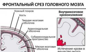
To understand what subarachnoid cerebral hemorrhage is, a short excursion into physiology, namely into the structure of the hemispheres, will help. Physiologically, the meninges consist of three balls:
- external, solid configuration;
- medium, spider type;
- internal, which is the vascular cover.
There is space between all the balls: the area between the first two balls is called subdural, and the area between the choroid and tunica media is called subarachnoid.
In a normal state, all membranes have an integral structure, which ensures protection of the hemispheres and normal brain activity. A case in which, due to difficulties in blood circulation, vascular spasms or traumatic events, an outpouring of blood occurs in the subarachnoid zone is identified as subarachnoid. Subarachnoid hemorrhage, abbreviated as SAH, may also be referred to as intracranial hemorrhage or stroke.
Hemorrhage of the subarachnoid type is often characterized by spontaneity, occurs against the background of a segmental or large-scale rupture of cerebral blood lines, and is accompanied by sharp and intense headaches, bouts of vomiting, and loss of consciousness. This is a very dangerous condition, often causing sudden death for the patient, and the chances of saving a person directly depend on the promptness of first aid and the intensity of blood filling in the subarachnoid zone.

Causes of effusion
Help for the progression of pathology is a violation of the tightness of the walls of the vascular highways of the hemispheres. The causes of subarachnoid hemorrhage can have different etiologies, mainly the following:
- Complex head injuries, which are accompanied by traumatic brain injuries, brain contusions or direct rupture of arteries in the hemispheres.
- An unexpected rupture of the artery wall, which can be caused by infectious diseases, a rapid increase in pressure, or may also occur due to the use of alcoholic beverages or drugs.
- Vascular malformation deformity.
Symptoms of pathology
Often, the progression of the pathology begins to make itself felt to a person with unpleasant symptoms, with its etiology of a neuralgic nature, several days before the onset of a massive outpouring. During this period, a characteristic feature is the thinning of the vessel wall, through which blood begins to leak in small volumes. This condition is accompanied by nausea and dizziness, visual impairment. In the absence of timely diagnosis and adequate treatment, the disease progresses, one or more vessels rupture, and blood begins to intensively fill the subarachnoid segments of the brain. Similar symptoms may be accompanied by traumatic subarachnoid hemorrhage if the head injury is not particularly intense.
Symptoms of extensive bleeding are pronounced, accompanied by sharp, explosive, diffuse pain in the head, followed by irradiation to the shoulders, neck and occipital region. Subarachnoid hemorrhage in the brain of a progressive type is often accompanied by nausea with bouts of vomiting, photophobia, disturbances of consciousness, often with fainting precedents and coma. The period from the onset of massive effusion to coma can range from several minutes to half a day.
In newborns, subarachnoid hemorrhage is predominantly a consequence of trauma during childbirth and is characterized by the formation of hematomas in the hemispheres. Cerebral hemorrhage in newborns is accompanied by the following symptoms:
- shrill, intense crying of a child against the background of increased physical activity;
- convulsive attacks;
- lack of sleep;
- involuntary eye movement, visual strabismus;
- extreme severity of innate reflexes;
- increased muscle tone;
- convexity of the fontanel with intense pulsation;
- jaundiced body color.

Symptoms of the pathology in a newborn can appear either immediately after birth or within several days, depending on the scale of the effusion in the hemispheres. If the problem is identified in a timely manner, modern medicine allows the child to be resuscitated, in most cases without negative consequences for his future life.
Prevalence of the disease and stages of its progression
Precedents associated with SAH of the brain are a fairly common phenomenon. According to statistics, the most common cases are considered to be cases of subarachnoid effusion due to trauma, accounting for about sixty percent of all cases.
Less common are precedents for the development of pathology due to changes in blood circulation in the cerebral vessels, diagnosed in seven percent of patients with this pathology. Most often these are patients of advanced and retirement age, as well as people with alcohol or drug addiction. The rarest cases are cases of spontaneous progression of the disease, their prevalence is less than one percent.
As for the etiology of the disease, the most common situations in medical practice are the occurrence of SAH due to rupture of arteries located in the circle of Visilli. Such precedents account for about eighty-five percent of all registered cases, half of them end in death, while fifteen percent of patients do not even have time to get to a medical facility.
Cerebral hemorrhage is a disease that most often affects the adult population, however, the pediatric category is no exception. In children, this pathology often occurs due to trauma. Subarachnoid hemorrhage in newborns can be the result of a prolonged or too rapid natural labor, when there is a mismatch between the mother’s birth canal and the baby’s head, as well as a consequence of the baby being without oxygen for a long time. The progression of pathology in a child can be provoked by infectious diseases of the mother, pathologies of brain activity in a child of the congenital category, and fetal hypoxia.

Medicine classifies SAH of traumatic origin into three stages of development:
- Progression of intracranial hypertension against the background of mixing of gushing blood with cerebrospinal fluid, increasing the latter in volume.
- An increase in hemispheric hypertension to extreme maximums, due to the formation of blood clots in the cerebrospinal fluid channels, their blocking and disturbances in the circulation of cerebrospinal fluid.
- Dissolution of blood clots, followed by intensification of inflammatory processes in the hemispheres.
Classification of disease severity
To assess the severity of a patient’s condition, medical specialists use three methodologies for ranking the course of pathology.
Most often in practice, the Hunt-Hess scale is used to categorize the patient’s condition, which has five degrees of damage to the human brain:
- The first degree of the disease is considered the least life-threatening if therapy is started in a timely manner, and is characterized by a high percentage of patient survival. At this stage, the disease is asymptomatic with minor headaches and the onset of stiffness of the neck muscles.
- The second degree of the disease is characterized by a distinct loss of mobility of the occipital muscles, intense headaches, and paresis of the nerves of the hemispheres. The prospects for a favorable outcome do not exceed sixty percent.
- The third degree of the disease manifests itself in a person as a moderate deficiency of the neuralgic category, stunning. The patient's chance of survival does not exceed fifty percent.
- The fourth level of pathology is characterized by the patient’s frozen state, and a first-degree coma may occur. Typical for this stage are failures of the autonomic system and severe hemiparesis. Chances of life are about twenty percent.
- Last degree of progression: second or third level coma. The prognosis for the patient is disappointing, survival rate is no more than ten percent.
The second, no less popular in medical practice for assessing a patient’s condition, is the Fisher gradation, which is based on the results of computed tomography:
- If a CT examination does not visually detect bleeding, the disease is assigned the first degree of severity.
- The second stage is assigned to pathology if the scale of the effusion does not exceed one millimeter in thickness.
- If the lesion is more than one millimeter in size, the third level of pathology progression is diagnosed.
- When blood spreads inside the ventricles and in the parenchyma, the fourth degree of progression of SAH is diagnosed.

The SAH severity scale according to the World Federation of Neurosurgeons ranks the disease as follows:
- The first stage is fifteen points on the GCS, no neurological deficit.
- The second level is from thirteen to fourteen points, with the absence of neurological impairment.
- Third level – scores are similar to the previous version, with signs of disorders in the nervous and peripheral systems.
- The fourth stage of progression is assigned from seven to twelve points on the Glasgow Coma Scale.
- The last stage of the disease: less than seven points were diagnosed according to the GCS.
Diagnosis of pathology
Subarachnoid hemorrhage belongs to the category of the most complex and life-threatening cases. Its diagnosis involves conducting a complex of hardware examinations of the patient in order to confirm the diagnosis, as well as determine the stage of development, localization of hemorrhage, and the degree of disorders in the vascular system and hemispheres.
The main examination procedures include:
- Initial examination of the patient, analysis of his complaints.
- Visual assessment of a person’s condition, monitoring of his consciousness and the presence of neurological abnormalities.
- A laboratory blood test that can be used to determine the criteria for blood clotting.
- Cerebrospinal fluid puncture. If about twelve hours have passed since the onset of hemorrhage, its results, namely the presence of blood in the cerebrospinal fluid, can confirm the progression of SAH.
- or computed tomography allows you to identify the presence and location of the effusion, as well as assess the general condition of the brain. CT is more informative in the situation with SAH, which is why this type of examination is often prescribed to patients.
- If there is a suspicion of brain displacement as a result of injury, echoencephalography is prescribed to confirm or refute this fact.
- Transcranial Doppler ultrasound is performed to monitor the quality of blood flow in the cerebral arteries and its deterioration as a result of narrowing of the blood vessels.
- Magnetic resonance angiography of the arteries helps to assess their integrity and patency.

Based on the results of the study, the patient will be diagnosed in accordance with the International Classification of Diseases, Tenth Revision. SAH is classified in the section “Diseases of the circulatory system,” a subgroup of cerebrovascular diseases, and may have an ICD-10 code from I160.0 to I160.9, depending on the location of the source of the effusion.
Treatment methods
The methodology for treating pathology involves both drug treatment and surgical intervention, depending on the stage of the disease and its complexity. The feasibility of therapy and its direction can only be determined by a qualified specialist solely on the basis of diagnostic results. Primary measures should be focused on stopping bleeding, stabilizing, preventing or reducing the volume of brain swelling.
First aid
First aid for subarachnoid hemorrhage does not provide for any specific procedures; it consists of immediately calling an ambulance. It is strictly forbidden to give the patient any medications to eliminate symptoms, as this can cause unpredictable consequences.
If a sick person has an epileptic seizure, you must try to create comfortable conditions for him by placing soft things under his head and other parts of the body. After the seizure ends, you need to lay the sick person on his side, try to fix his limbs and wait for the ambulance to arrive.
When a person is unconscious as a result of cardiac arrest, it is necessary to perform cardiopulmonary resuscitation, with the proportion of chest compressions to breaths being thirty to two.
When there is an outpouring into the hemispheres, the only rational help for the patient is his hospitalization as soon as possible. All restorative and therapeutic procedures are subsequently carried out exclusively under the guidance of specialists, based on the results of diagnosing the patient’s condition.
Drug treatment
Conservative therapy can be used in situations where there are no indicators for surgical intervention, as well as to normalize the patient’s condition in the preoperative and postoperative period.
The main objectives of drug treatment of subarachnoid hemorrhage are:
- achieving stability of the patient's condition;
- prevention of relapses;
- stabilization of homeostasis;
- eliminating the original source of the outpouring;
- carrying out treatment and preventive measures aimed at prevention.
Depending on the complexity of the disease and its manifestations, the patient may be prescribed the following medications:

The appropriateness, dosage and duration of taking medications are determined exclusively by the attending physician and are based on medical indicators. During the treatment process, the doctor monitors the dynamics and can change the quantitative and qualitative composition of the drugs if there are no positive results.
Surgery
Surgical intervention is often prescribed by medicine for existing intracranial hematomas of significant size or when SAH occurs as a result of a serious head injury. In a situation where the patient experiences massive bleeding, emergency surgical procedures are performed. In other cases, the timing of the operation may vary and depend on the condition and age of the patient, the volume of effusion and the complexity of the symptoms.
Medicine provides the following types of surgical intervention for subarachnoid effusion:
- Removal of hemorrhagic contents by inserting a syringe or a specific needle.
- Elimination of hematoma with opening of the skull.
- Laser coagulation of blood vessels, if the effusion cannot be stopped with medications, sometimes with the application of specific clips to the damaged areas of the artery.
After surgery, the patient will have to undergo a mandatory course of drug therapy.
Rehabilitation procedures
Measures to restore the patient after subarachnoid hemorrhage are a mandatory continuation of therapy in the postoperative period. Depending on the complexity of the illness suffered, rehabilitation can last from six months to several years and has a complex structure.
After the incident, it is important for the patient to completely abandon bad habits, try to avoid stressful situations and maintain a healthy lifestyle. In addition, during the rehabilitation period, medicine provides for the use of medications, the action of which is aimed at preventing relapses.
Rehabilitation of the patient, depending on the severity of the illness experienced, may include the following areas:
- specific massages and hardware procedures to restore the patient’s muscle and motor activity;
- health treatments in special centers;
- therapeutic exercises to restore walking and coordination skills;
- classes with a psychologist to restore the patient’s psycho-emotional state.

During the recovery process at home, the patient will need proper care, as well as the support of loved ones.
Prognosis and possible complications
Subarachnoid cerebral hemorrhage is an insidious disease that very rarely goes away without a trace for a person. The most harmless complications are in the form of frequent migraines and disturbances in hormonal regulation of the body. Additionally, after experiencing an illness, the patient may experience a deterioration in brain activity, manifested in the form of psycho-emotional disorders, deterioration of attention and memory. However, such manifestations of the body after SAH are not considered particularly dangerous. Dangerous consequences include:
- vasospasm, which often provokes ischemic processes in the hemispheres;
- delayed ischemia, which affects more than a third of all patients, entails irreversible brain starvation with all the ensuing consequences;
- recurrent exacerbation of pathology;
- hydrocephalus;
- Rare complications include pulmonary edema and heart attacks.
The chances of a patient’s recovery after SAH depend on many factors, such as the person’s general physical health, his age, the stage of the disease and the extent of the effusion, and the promptness of first aid.
Often, it is a belated visit to a medical institution against the backdrop of a heavy outpouring that causes death for the patient or serious complications that do not allow the person to return his life to normal.
Preventive measures
Prevention of SAH, like many other diseases of the cardiovascular system, is not particularly difficult. The main rule, the observance of which helps to prevent cerebral hemorrhage, in addition to precedents with injuries, is a healthy lifestyle. A balanced diet, giving up bad habits, regular walks in the fresh air and moderate physical activity to keep the body in excellent condition, timely treatment of problems with blood vessels and heart under the supervision of doctors are the primary and effective preventive measures against the development of SAH and other complex ailments.
If a person has prerequisites for the development of SAH caused by cardiac problems, it is worth undergoing regular examinations, taking preventive medications prescribed by doctors as necessary to normalize blood pressure and heart rate, and monitor the state of one’s health.
In this case, careful attention to your body and a correct lifestyle are the most important preventive measures that help to avoid a complex and life-threatening incident.
Let's sum it up
Subarachnoid hemorrhage belongs to the category of the most dangerous diseases, which very often cause death. Of course, it is better to prevent such situations, however, if such a precedent occurs, it is worthwhile to immediately deliver the patient to a medical facility: a person’s life depends on the speed of diagnosis and provision of correct assistance.
Lead a full, healthy and correct lifestyle - this will help you avoid many health problems, is the key to the proper functioning of the body, and reduces the risk of developing not only SAH, but also other diseases.