What is feces on I. Analysis of feces for helminth eggs (helminth eggs)
The portal contains information about private clinics and medical centers in St. Petersburg where you can take a coprogram - general analysis feces To make it more convenient to choose a medical institution, we have prepared a special conditional rating, collected patient reviews and indicated in tables the prices for the most popular medical and diagnostic procedures.
Visitors to the portal will be able to quickly choose where to take stool tests in St. Petersburg based not only on the cost of the procedure, but also on reviews of the work of the medical center’s specialists. Depending on the laboratory, the results and transcript of the stool analysis can be obtained both on the day the material is submitted, and after 6 days.
A stool coprogram is a special analysis aimed at studying the physical, chemical and microscopic properties of material taken from the patient. By performing the procedure for preventive purposes, it is possible to promptly identify abnormalities in the functioning of the digestive system and diagnose dangerous diseases. Feces are the final result of the activity of the digestive organs, clearly reflecting the nuances of the functioning of each individual organ.
How should stool be collected for analysis in the laboratory?
Material for research should be collected after spontaneous bowel movement in special disposable containers, which can be purchased at a pharmacy or obtained in the treatment room. The amount of feces should not exceed one third of the volume of the entire container. It will require you to indicate the patient’s name, date of birth, time and date of collection of the material.
Stool analysis will not give a reliable result on material collected after taking medications, after an enema, taking vaseline or castor oil, administering suppositories or preparations with bismuth, iron or barium sulfate. There should be no urine impurities in the stool. The resulting biomaterial must be delivered to the laboratory no later than 10 hours after its collection if it was stored at a temperature of 4 - 8 degrees.
Is any specific patient preparation required?
 Carrying out a scatological examination does not require preliminary preparation of the patient. The most you need to remember is the need to temporarily stop taking certain medications that affect the appearance of stool and increase intestinal motility.
When is a stool test for coprogram prescribed?
The procedure is prescribed by a doctor in order to identify diseases of the digestive organs, as well as to evaluate the effectiveness of the treatment already prescribed by the doctor. Carrying out a coprogram will be a useful action in diagnosing diseases and pathological conditions of the body such as:
- Stomach ulcers, allergic colitis and other similar diseases.
- Excessively accelerated passage of food through the intestines.
- Pronounced inflammation located in the area gastrointestinal tract.
- Presence of violations in duodenum complicating absorption.
- Problems in the functioning of the stomach, liver or pancreas.
- Other pathological abnormalities.
This type of diagnostics takes a minimum amount of time, allowing you to quickly detect existing deviations. Thus, the doctor promptly determines the optimal treatment for the patient’s health.
Food, passing through the gastrointestinal tract, undergoes successive transformations and is gradually absorbed. Feces are the result of the digestive system. When examining feces, the condition of the digestive system and various digestion defects are assessed. Therefore, scatology is an indispensable component in the diagnosis of diseases of the gastrointestinal tract and helminthiases.
There are different types of stool examinations. Which of them will be done is determined by the purpose of the study. This can be a diagnosis of gastrointestinal pathology, helminthiases, and changes in microflora. Clinical analysis of stool is sometimes carried out selectively, only according to the parameters necessary in a particular case.
General analysis
Examination of excrement can be divided into general stool analysis and examination under a microscope (called a coprogram). In general, the quantity, smell, color, consistency, impurities are examined; microscopic analysis reveals undigested muscle and plant fibers, salts, acids and other inclusions. Nowadays, a coprogram is often called general analysis. Thus, CPG is the study of physical, chemical properties feces and pathological components in them.
Stool tests to detect protozoa are carried out if amoebiasis or trichomoniasis is suspected. Trichomonas are difficult to see in feces. When taking material for this purpose, you cannot use enemas, laxatives, or treat the feces container with disinfectant liquids. The interpretation will be correct only if examined immediately, a maximum of 15 minutes after collecting the material. The search for cysts does not require such urgency; they are resistant to external environment. To reliably detect Shigella, a piece of feces with blood or mucus is taken and placed in a container with a special preservative.
Clinical picture
Make an appointment with a doctor right now!
Doctor of Medical Sciences, Professor Morozova E.A.:

Get tested>>
Stool analysis shows the presence of pathogens in the body intestinal infections and ratio various types bacteria.
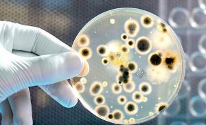
Sowing on nutrient media will make it possible to objectify quantitative and qualitative changes in the intestinal microflora.
Stool analysis should be carried out no later than three hours after taking the morning portion of stool. It is advisable to store the sample refrigerated (). Stool analysis cannot be performed during antibiotic therapy, optimally two weeks after its completion. It is important to avoid urine and vaginal discharge, especially during menstruation. The volume of the sample should be at least 10 ml, the sample should be taken from different parts of the feces, making sure to include areas with mucus and blood.
Analysis of stool by scraping in the perianal area is carried out to detect pinworm eggs. The material must be examined no later than three hours after collection.
So what does the analysis show:
- protozoa and microbes that cause intestinal infections;
- the presence of helminths and their eggs;
- state of microflora;
- digestion defects;
- effectiveness of treatment (with follow-up);
- in children - signs of cystic fibrosis and lactose deficiency.
Rules for conducting research
To obtain reliable data, you need to know how to properly collect feces and when a stool analysis should be deciphered.
An example of a correctly taken sample:
- Before the examination, you should be on a diet for several days that excludes flatulence, stained stools, retention or diarrhea.
- A scatological analysis of stool should be taken during natural bowel movements. Enemas, laxatives, including rectal suppositories, Microlax microenemas cannot be used, as the true picture of the study may be distorted.
- A general stool analysis is reliable if, within three days before collecting the material, the patient did not take medications that could change the color or character of feces (barium, iron, bismuth).
- A scatological analysis of stool should be carried out no later than five hours after collecting the material.
- The optimal volume for testing is about two teaspoons (about 30 grams of feces).
- To detect helminthiasis, it is better to take samples from different parts of a portion of excrement.
- The material should be collected in a sterile container.
Decoding the research results
 It is very important to correctly decipher the stool analysis. To do this, you need to know the research algorithm and normal indicators.
The patient's decoding includes three main points: macroscopy (examination), biochemistry, microscopy (the actual coprogram).
Inspection
Clinical analysis of stool begins with its visual assessment. The norm implies a dense consistency and dark color of excrement, the absence of mucus, blood, foul odor, undigested food particles and other pathological impurities.
Biochemistry
A chemical analysis of stool is performed.
A normal stool analysis implies the following negative biochemical reactions to the following elements:
- occult blood;
- bilirubin;
- iodophilic flora;
- starch;
- protein;
- fatty acid.
The reaction to stercobilin should be positive (75–350 mg per day). It provides color and reflects the functioning of the liver and large intestine; its amount increases with hemolytic anemia and decreases with disorders of the outflow of bile.
Ammonia is normally 20–40 mmol/kg.
It is important to determine the acid-base state of excrement using litmus paper; the pH of feces should be close to neutral values (6-8). Changes in the acidity of intestinal contents are possible due to microflora or diet disorders.
Microscopy
A stool analysis under a microscope is also necessary. The coprogram carries information about the presence of pathological components in excrement and allows one to assess the quality of food digestion. Examination of stool in children will help in the diagnosis of infections and inflammation of the gastrointestinal tract, cystic fibrosis, enzymatic and dysbacterial disorders, helminthic infestations.
The norm implies the absence of the following substances:
- undigested fat and its derivatives;
- muscle fibers;
- connective tissue;
- crystals from the remains of destroyed blood cells.
Yeast and other fungi are also normally absent in stool analysis.
Stool microscopy is also used to objectively assess the dynamics of the patient’s condition.
What diseases can a stool test help diagnose?
What do certain deviations from the norm that were found when laboratory research excrement? Options for changing normal stool parameters exist for various diseases.
Deviations during macroscopy
Discoloration indicates gallstone disease, since the stones disrupt the flow of bile, stercobilin does not enter the intestines, and the stool loses its dark color. This phenomenon is observed in pancreatic cancer, hepatitis, and cirrhosis of the liver.
Black color, tar consistency - a sign peptic ulcer, a tumor complicated by gastric bleeding.
Reddish color of stool causes bleeding in the lower intestines.
The foul odor is due to rotting or fermentation in the gastrointestinal tract. Its appearance is possible when chronic pancreatitis, dysbacteriosis, cancer.
Elements of undigested food may be found in the excrement. This indicates a deficiency of gastric juice, bile, enzymes, or an acceleration of peristalsis when food simply does not have time to be absorbed.
Fresh blood is possible for anal fissures, hemorrhoids, ulcerative colitis
Mucus plays a protective role. Its detection indicates the presence of inflammation of the intestinal walls. , dysentery, colitis are characterized by a large amount of mucus in the excrement. Mucus is also found in cystic fibrosis, celiac disease, malabsorption syndromes, irritable bowel syndrome, hemorrhoids, and polyps.
Changes in biochemistry
If there is a change in the acid-base properties of the stool being tested, this indicates disturbances in the digestion of food. The alkaline environment of excrement is a consequence of putrefactive processes due to impaired protein breakdown, the acidic environment is due to fermentation, which is observed with excess consumption or impaired absorption of carbohydrates.
Occult blood testing is used to detect gastric and intestinal bleeding in peptic ulcers, polyps, cancer of various parts of the gastrointestinal tract, and the presence of helminths. To avoid erroneous results, three days before the intended collection of material, foods containing iron should be excluded from the diet, and traumatic procedures such as FGDS and colonoscopy should not be performed. If you have periodontal disease, it is better not to brush your teeth on the day of the test so that there is no blood from the diseased gums.
Bilirubin can be detected when acute poisoning, gastroenteritis.
Protein is found in pancreatitis and atrophic gastritis.
If starch appears, it is necessary to exclude pancreatitis, malabsorption, and pathology of the small intestine.
Iodophilic flora appears in dysbacteriosis, pathology of the pancreas, stomach, and fermentative dyspepsia. They are especially often found during fermentation, acid reaction of intestinal contents and acceleration of its evacuation.
Ammonia increases during putrefactive processes, against the background of inflammation and impaired protein digestion.
Deviations of microscopic analysis
A lot of muscle fibers in excrement are observed with pancreatitis and atrophic gastritis. They can be found in young children, with diarrhea, and poor chewing of tough meat.
Connective fibers can be found in gastritis with low acidity, pancreatitis, when eating poorly cooked meat.
If neutral fat is detected, the elements fatty acids and their salts, this indicates insufficient production of bile and pancreatic enzymes. Possible reasons:
- pancreatitis;
- pancreatic tumor;
- stones in the bile ducts;
- increased peristalsis when fats do not have time to be absorbed;
- malabsorption in the intestine;
- eating too fatty foods;
- use of rectal suppositories.
In children, the presence of fat may be associated with incompletely developed digestive function.
When the acidity of excrement changes to the alkaline side, soaps (salts of undigested fatty acids) are found. Their detection in large quantities in adults is possible due to accelerated peristalsis and pathology of the biliary tract.
Soluble plant fiber indicates decreased production of gastric juice and other enzymes.
Appearance yeast-like fungi speaks of dysbiosis due to immunodeficiency or antibiotic therapy.
In stool analysis, a high level of leukocytes is noted in cases of inflammation in the gastrointestinal tract, rectal fissures, and oncology.
How to prepare for scraping for enterobiasis?
There should be no specific preparations before scraping for enterobiasis. The only recommendation is do not carry out toilet the anal area and do not go to the toilet “too much”, this will allow you to get the most reliable analysis results.How is scraping performed for enterobiasis?
- The material is collected in the morning immediately after you wake up. (do not toilet the anal area and do not go to the toilet “in large quantities”)
- You can collect the material in a laboratory or clinic/hospital. (This best option, since you won’t have to rack your brains about how and what to do, and will also allow you to get the most recent material for research.)
- If you decide to collect the material in person, then the material must be collected in a special plastic tube or on a glass slide with a sticker.
How to collect material in a plastic container?
How to collect material on a special glass slide with transparent adhesive tape pasted on top?
FAQ
How to prepare for a stool test for helminth eggs?
In fact, there should be no specific preparations before taking a stool test for helminth eggs.How to properly collect stool for analysis for helminth eggs?
- Urinate before collecting material to prevent urine from getting into the stool.
- It is necessary to take a clean, dry container where defecation will be carried out.
- From the resulting material you need to take 8-10 cm 3 (~2 teaspoons). Feces are collected using a special “spoon” that is built into the lid of a special container that you should be given to collect feces. Read more about the technique by following the link: Collecting material in a container.
- Feces for analysis are collected from different parts of the stool (top, sides, inside)
- The material (feces) is placed in the container given to you and closed tightly.
- You must sign the container (your first and last name, date of analysis collection)
Is it possible to store feces for analysis for helminth eggs?
To obtain the most accurate data, it is strongly recommended to submit stool for analysis for helminth eggs within 30-45 minutes after defecation.You can store feces for 5-8 hours in the refrigerator, in a tightly closed container at a temperature of +4ºС-+8ºС. However, storage may adversely affect test results.
How much stool should be collected for analysis for helminth eggs?
For analysis, it is necessary to collect 8-10 cm 3 (~2 teaspoons) of feces.Is it possible to store the obtained material for analysis for enterobiasis?
It is recommended to take the resulting material to the laboratory as soon as possible, this will allow for the highest quality analysis and will allow obtaining the most reliable results. However, if you do not have the opportunity to take the material to the laboratory in the near future, you can store the material in the refrigerator at a temperature of +4ºС / +8ºС for 8 hours. The longer the material remains in the refrigerator, the less accurate the results will be. Not all people know how to properly collect stool samples. This is especially important when a coprogram is done; stool analysis will not be informative if the stool is collected incorrectly. Stool after using suppositories or performing enemas is not suitable for research. You should not use medications for constipation before donating stool. A few days before the test, the patient must exclude all drugs that could somehow affect the functioning of the gastrointestinal tract. You should not take iron supplements if your stool is being tested. Typically, a study is prescribed when there is bloating, diarrhea and constipation, stools of questionable color or consistency, abdominal pain and other symptoms.
Analysis of feces for worms. Such a study is often prescribed to children. When registration is required for educational and other institutions and for the purpose of prevention. Worms occur frequently in children. This analysis does not require special preparation. Normally, there should be no worm eggs in the stool.
Analysis of stool for dysbacteriosis. This study helps to identify microflora disorders. Doctors check the content of bifidobacteria, lactobacilli, pathogenic bacteria and fungi. The presence of bacteria such as staphylococci, salmonella, clostridia, enterobacteria, coli and other pathogenic bacteria. Bacteria can be opportunistic and pathogenic. Normally, they should not be present in the stool at all. Opportunistic bacteria may be present in small quantities. Frequent bowel movements are usually an indication for testing.
Test for blood present in stool. If doctors suspect bleeding in a person’s gastrointestinal tract, the presence of ulcers, or polyps, then an occult blood test is done. Sometimes it becomes the first diagnostic test to detect colorectal cancer. Preparation for this test is the same as for other stool tests, but the patient must follow a diet. Iron supplements and any meat are excluded 3 days before the test.
How to properly prepare for a stool test
Stool collection is carried out in the evening or in the morning before delivery to the laboratory. The jar must be clean and dry. The child takes feces from the diaper or diaper, or from the potty. If stool is collected in the evening, it is placed in a jar with a tight-fitting lid and stored in the refrigerator. Feces can be stored for no more than 12 hours. Here you can take a stool test, the price of which is very reasonable. Contact us to receive timely and most reliable analysis. It is very important to undergo a timely examination in order to promptly detect serious diseases that require treatment.
Make an appointment
Food, passing through the human intestinal tract, is subject to gradual transformations, gradually being absorbed. Feces are the result of the digestive system. During the examination of feces, the condition of the organs of the digestive system and various digestive disorders are determined. Therefore, scatology is an obligatory element in the diagnosis of helminthiasis and gastrointestinal diseases.
Eat different types taking a stool test. What examinations will be performed are determined by the main purpose of donating stool. This makes it possible to diagnose changes in microflora, helminthiases, gastrointestinal diseases, etc. Clinical analysis of stool in some cases is carried out selectively, only according to the necessary parameters in a particular case.
General analysis
Examination of excrement is divided into examination under a microscope ( coprogram) And general stool analysis. During the general procedure, the smell, quantity, impurities, consistency, color are examined, the coprogram determines undigested acids, salts, vegetable and muscle fibers and other inclusions. Today, a coprogram is often called general analysis.
 Stool tests for protozoa are performed when there is a suspicion of trichomoniasis or amoebiasis. It is difficult to see Trichomonas in feces. When collecting material for this purpose, it is prohibited to treat containers for feces with disinfectants, use laxatives, or enemas. The interpretation is correct only with immediate examination of no more than 15 minutes after collecting material.
The identification of Giardia cysts does not require this urgency; they are characterized by persistence in the external environment. To reliably determine Shigella, a fragment of feces is taken with mucus or blood and placed in a test tube with a special preservative.
Bacteriological analysis of stool determines the presence of pathogens of intestinal infections in the body and the ratio of different types of bacteria.
Sowing on nutrient media will make it possible to objectify qualitative and quantitative changes in the intestinal microflora.
 Bacteriological analysis of stool must be performed no later than 3 hours after collecting stool in the morning. It is best to keep the sample refrigerated. This stool test prohibited during antibiotic therapy, preferably 10 days after its end. Necessary exclude hit vaginal discharge and urine. The volume of the sample must be at least 10 ml; collection should be done from various areas of feces, always including areas with blood and mucus.
A scraping in the perianal area is performed to identify pinworm eggs. The material must be examined no later than 3 hours after collection.
So, what will a stool analysis show:
- the presence of helminths and their eggs;
- microbes and protozoa that cause intestinal infections;
- digestive disorders;
- state of microflora;
- in children - signs of insufficiency of lactose synthesis and cystic fibrosis;
- effectiveness of treatment.
Rules for performing the examination
To have reliable data, you need to know how to collect feces and when the analysis must be deciphered.
How to properly take a stool sample:

Decoding survey data
Quite important to do correct decoding stool analysis. Why do you need to know the normal indicators and examination algorithm?
The transcript includes three main points: examination, biochemistry, coprogram (microscopy).
Inspection
Clinical analysis of the composition occurs with its visual assessment. The norm implies dark color of excrement, dense consistency, absence of blood, mucus, undigested food particles, fetid odor and other pathological conditions.
Biochemistry
A chemical analysis of feces is performed.
Normal analysis shows negative biochemical reactions for the following elements:
- bilirubin;
- occult blood;
- starch;
- iodophilic microflora;
- fatty acid;
- protein.
 The reaction to stercobilin must be positive. It reflects the work of the large intestine and liver, and also provides color; its amount is reduced in case of bile outflow disorders, and increased in hemolytic anemia. It is important to identify the acid-base state of feces using litmus paper; the pH of feces should be close to neutral (6-8). Changes in acidity are possible due to diets or microflora disorders.
Microscopy
A fecal examination under a microscope is also required. The coprogram determines the presence of pathological impurities in excrement and makes it possible to assess the quality of digestion. Examination of stool in children can help in the diagnosis of dysbiosis, gastrointestinal inflammation and infections, cystic fibrosis, helminthic infestations, dysbacterial and enzymatic disorders.
Normally they should such substances are absent:
- muscle fibers;
- undigested fat and its derivatives;
- crystals from particles of destroyed blood cells;
- connective tissue.
Yeasts and other fungi are also normally absent.
What do specific deviations from the norm that were discovered during a laboratory examination of stool indicate? There are options for changing acceptable stool levels for various diseases.
Deviations during macroscopy:
- The tar consistency and black color are symptoms of a peptic ulcer, a tumor that is complicated by gastric bleeding.
- Discoloration indicates cholelithiasis, since stercobilin does not pass into the intestines, stones interfere with the flow of bile, and feces lose their dark hue. This phenomenon is observed in liver cirrhosis, hepatitis, and pancreatic cancer.
- The foul odor is caused by fermentation or rotting in the gastrointestinal tract. Its manifestation can be in cancer, dysbiosis in children, chronic pancreatitis.
- The reddish color of the stool creates bleeding in the lower intestinal tract.
- Mucus has a protective function. Its definition indicates the presence of an inflammatory process of the intestinal walls. Colitis, dysentery, and salmonellosis are characterized by a significant amount of mucus in excrement.
- Fresh blood can be found in anal fissures, dysentery, ulcerative colitis, and hemorrhoids.
- Particles of undigested food may be found in the stool. This indicates a deficiency of enzymes, bile, gastric juice, or an acceleration of peristalsis, in which case the food does not have time to be absorbed.
Changes during biochemistry:
- An examination for occult blood is used to determine intestinal and gastric bleeding in polyps, peptic ulcers, the presence of helminths, and cancer of various parts of the gastrointestinal tract. To avoid erroneous results for 3 days, before donating material, foods that contain iron must be excluded from the diet, and traumatic procedures such as colonoscopy and FGDS are prohibited. During periodontal disease, on the day when it is necessary to take the test, you should not brush your teeth, this will eliminate blood impurities from the infected gums.
- When there is a change in the acid-base parameters of the examined feces, this indicates a digestive disorder. The alkaline environment of feces is the result of putrefactive processes during a violation of protein breakdown, acidic - during fermentation, this occurs when absorption is impaired or excessive consumption of carbohydrates.
- Protein is found in atrophic gastritis and pancreatitis.
- Bilirubin can be detected in gastroenteritis and acute poisoning.
- Iodophilic microflora appears in dysbacteriosis in children, fermentative dyspepsia, pathology of the stomach and pancreas.
- When starch appears, it is necessary to exclude pathology of the small intestine, malabsorption, and pancreatitis.
Deviations during microscopic examination:

When the elements of fatty acids and salt derivatives, neutral fat are determined, this indicates insufficient production of enzymes and bile in the pancreas. Probable reasons:
- pancreatic oncology;
- pancreatitis;
- increased peristalsis;
- stones in the bile ducts;
- consumption of very fatty foods;
- use of rectal suppositories;
- intestinal absorption disorder.
You need to know why donate feces and what the purpose of the study is. The delivery of feces must be carried out in accordance with all the rules, observing the nuances that are characteristic of specific tests. The examination must be taken seriously if you want to get accurate results, correct diagnosis of the disease and adequate treatment.
