Human musculoskeletal system. ODS hygiene
In the process of evolution, animals mastered more and more new territories, types of food, and adapted to changing living conditions. Evolution gradually changed the appearance of animals. In order to survive, it was necessary to search for food more actively, hide better or defend against enemies, and move faster. Changing along with the body, the musculoskeletal system had to ensure all these evolutionary changes. The most primitive protozoa have no supporting structures, move slowly, flowing with the help of pseudopods and constantly changing shape.
The first support structure to appear is cell membrane. It not only separated the organism from the external environment, but also made it possible to increase the speed of movement due to flagella and cilia. Multicellular animals have a wide variety of support structures and devices for movement. Appearance exoskeleton increased the speed of movement due to the development of specialized muscle groups. Internal skeleton grows with the animal and allows it to reach record speeds. All chordates have an internal skeleton. Despite significant differences in the structure of musculoskeletal structures in different animals, their skeletons perform similar functions: support, protection of internal organs, movement of the body in space. The movements of vertebrates are carried out due to the muscles of the limbs, which carry out such types of movement as running, jumping, swimming, flying, climbing, etc.
Skeleton and muscles
The musculoskeletal system is represented by bones, muscles, tendons, ligaments and other connective tissue elements. The skeleton determines the shape of the body and, together with the muscles, protects the internal organs from all kinds of damage. Thanks to joints, bones can move relative to each other. The movement of bones occurs as a result of contraction of the muscles that are attached to them. In this case, the skeleton is a passive part of the motor apparatus that performs a mechanical function. The skeleton consists of dense tissues and protects internal organs and the brain, forming natural bone containers for them.
In addition to mechanical functions, the skeletal system performs a number of biological functions. Bones contain the main supply of minerals that are used by the body as needed. The bones contain red bone marrow, which produces blood cells.
The human skeleton includes a total of 206 bones - 85 paired and 36 unpaired.

Bone structure
Chemical composition of bones
All bones consist of organic and inorganic (mineral) substances and water, the mass of which reaches 20% of the mass of the bones. Organic matter of bones - ossein- has elastic properties and gives elasticity to bones. Minerals - salts of carbon dioxide and calcium phosphate - give bones hardness. High bone strength is ensured by a combination of the elasticity of ossein and the hardness of the mineral substance of bone tissue.

Macroscopic bone structure
On the outside, all bones are covered with a thin and dense film of connective tissue - periosteum. Only the heads of long bones do not have periosteum, but they are covered with cartilage. The periosteum contains many blood vessels and nerves. It provides nutrition to bone tissue and takes part in the growth of bone thickness. Thanks to the periosteum, broken bones heal.
Different bones have different structures. A long bone looks like a tube, the walls of which consist of a dense substance. This tubular structure long bones gives them strength and lightness. In the cavities of the tubular bones there is yellow bone marrow- loose connective tissue rich in fat.

The ends of the long bones contain cancellous bone substance. It also consists of bony plates that form many intersecting septa. In places where the bone is subject to the greatest mechanical load, the number of these partitions is highest. The spongy substance contains red bone marrow, the cells of which give rise to blood cells. Short and flat bones also have a spongy structure, only on the outside they are covered with a layer of damlike substance. The spongy structure gives bones strength and lightness.
Microscopic structure of bone
Bone tissue belongs to the connective tissue and has a lot of intercellular substance, consisting of ossein and mineral salts.

This substance forms bone plates arranged concentrically around microscopic tubules that run along the bone and contain blood vessels and nerves. Bone cells, and therefore bone, are living tissue; it receives nutrients from the blood, metabolism occurs in it, and structural changes can occur.

Types of bones
The structure of bones is determined by the process of long historical development, during which the body of our ancestors changed under the influence of the environment and adapted through natural selection to the conditions of existence.
Depending on the shape, there are tubular, spongy, flat and mixed bones.
Tubular bones are located in organs that make rapid and extensive movements. Among the tubular bones there are long bones (humerus, femur) and short bones (phalanxes of the fingers).
Tubular bones have a middle part - the body and two ends - the heads. Inside the long tubular bones there is a cavity filled with yellow bone marrow. The tubular structure determines the bone strength required by the body while requiring the least amount of material. During the period of bone growth, between the body and the head of the tubular bones there is cartilage, due to which the bone grows in length.
Flat bones They limit cavities within which organs are placed (skull bones) or serve as surfaces for muscle attachment (scapula). Flat bones, like short tubular bones, are predominantly composed of spongy substance. The ends of long tubular bones, as well as short tubular and flat bones, do not have cavities.

Spongy bones built primarily of spongy substance covered with a thin layer of compact. Among them, there are long spongy bones (sternum, ribs) and short ones (vertebrae, carpus, tarsus).
TO mixed bones These include bones that are made up of several parts that have different structures and functions (temporal bone).
Protrusions, ridges, and roughness on the bone are places where muscles are attached to the bones. The better they are expressed, the more developed the muscles attached to the bones are.
Human skeleton.
The human skeleton and most mammals have the same type of structure, consisting of the same sections and bones. But man differs from all animals in his ability to work and intelligence. This left a significant imprint on the structure of the skeleton. In particular, the volume of the human cranial cavity is much larger than that of any animal that has a body of the same size. The size of the facial part of the human skull is smaller than the brain, but in animals, on the contrary, it is much larger. This is due to the fact that in animals the jaws are an organ of defense and acquisition of food and are therefore well developed, and the volume of the brain is less than in humans.
The curves of the spine, associated with the movement of the center of gravity due to the vertical position of the body, help a person maintain balance and soften shocks. Animals do not have such bends.
The human chest is compressed from front to back and close to the spine. In animals it is compressed from the sides and extended towards the bottom.
The wide and massive human pelvic girdle has the shape of a bowl, supports the abdominal organs and transfers body weight to the lower limbs. In animals, body weight is evenly distributed between the four limbs and the pelvic girdle is long and narrow.
The bones of the lower limbs of humans are noticeably thicker than the upper ones. In animals there is no significant difference in the structure of the bones of the fore and hind limbs. Greater mobility of the forelimbs, especially the fingers, allows a person to perform a variety of movements and types of work with his hands.
Skeleton of the torso axial skeleton
Skeleton of the torso includes a spine consisting of five sections, and the thoracic vertebrae, ribs and sternum form chest(see table).

Scull
The skull is divided into the brain and facial sections. IN brain The section of the skull - the cranium - contains the brain, it protects the brain from blows, etc. The skull consists of fixedly connected flat bones: the frontal, two parietals, two temporal, occipital and sphenoid. The occipital bone is connected to the first vertebra of the spine using an ellipsoidal joint, which allows the head to tilt forward and to the side. The head rotates along with the first cervical vertebra due to the connection between the first and second cervical vertebrae. There is a hole in the occipital bone through which the brain connects to the spinal cord. The floor of the skull is formed by the main bone with numerous openings for nerves and blood vessels.

Facial the skull section forms six paired bones - the upper jaw, zygomatic, nasal, palatine, inferior nasal concha, as well as three unpaired bones - the lower jaw, vomer and hyoid bone. The mandibular bone is the only bone of the skull that is movably connected to the temporal bones. All bones of the skull (with the exception of the lower jaw) are connected motionlessly, which is due to their protective function.
The structure of the human facial skull is determined by the process of “humanization” of the monkey, i.e. the leading role of labor, the partial transfer of grasping function from the jaws to the hands, which have become organs of labor, the development of articulate speech, the consumption of artificially prepared food, which facilitates the work of the masticatory apparatus. The cranium develops in parallel with the development of the brain and sensory organs. Due to the increase in brain volume, the volume of the cranium has increased: in humans it is about 1500 cm 2.
Skeleton of the torso
The skeleton of the body consists of the spine and rib cage. Spine- the basis of the skeleton. It consists of 33–34 vertebrae, between which there are cartilage pads - discs, which gives the spine flexibility.
The human spinal column forms four curves. In the cervical and lumbar spine they are convexly facing forward, in the thoracic and sacral spine - backward. In the individual development of a person, bends appear gradually; in a newborn, the spine is almost straight. First, the cervical curve forms (when the child begins to hold his head straight), then the thoracic curve (when the child begins to sit). The appearance of lumbar and sacral curves is associated with maintaining balance in an upright position of the body (when the child begins to stand and walk). These bends have important physiological significance - they increase the size of the thoracic and pelvic cavities; make it easier for the body to maintain balance; soften shocks when walking, jumping, running.

With the help of intervertebral cartilage and ligaments, the spine forms a flexible and elastic column with mobility. It is not the same in different parts of the spine. The cervical and lumbar spine have greater mobility; the thoracic spine is less mobile, as it is connected to the ribs. The sacrum is completely motionless.

There are five sections in the spine (see diagram “Divisions of the spine”). The size of the vertebral bodies increases from the cervical to the lumbar due to the greater load on the underlying vertebrae. Each vertebrae consists of a body, a bony arch and several processes to which muscles are attached. There is an opening between the vertebral body and the arch. The foramina of all vertebrae form spinal canal where the spinal cord is located.
Rib cage formed by the sternum, twelve pairs of ribs and thoracic vertebrae. It serves as a container for important internal organs: heart, lungs, trachea, esophagus, large vessels and nerves. Takes part in respiratory movements due to the rhythmic raising and lowering of the ribs.
In humans, in connection with the transition to upright walking, the hand is freed from the function of movement and becomes an organ of labor, as a result of which the chest experiences a pull from the attached muscles of the upper limbs; the insides do not press on the front wall, but on the lower one, formed by the diaphragm. This causes the chest to become flat and wide.
Skeleton of the upper limb
Skeleton of the upper limbs consists of the shoulder girdle (scapula and collarbone) and the free upper limb. The scapula is a flat, triangular bone adjacent to the back of the rib cage. The collarbone has a curved shape, reminiscent of the Latin letter S. Its significance in the human body is that it sets the shoulder joint some distance from the chest, providing greater freedom of movement of the limb.
The bones of the free upper limb include the humerus, the bones of the forearm (radius and ulna) and the bones of the hand (bones of the wrist, bones of the metacarpus and phalanges of the fingers).

The forearm is represented by two bones - the ulna and the radius. Due to this, it is capable of not only flexion and extension, but also pronation - turning inward and outward. The ulna at the top of the forearm has a notch that connects to the trochlea of the humerus. The radius bone connects to the head of the humerus. In the lower part, the radius has the most massive end. It is she who, with the help of the articular surface, together with the bones of the wrist, takes part in the formation of the wrist joint. On the contrary, the end of the ulna here is thin, it has a lateral articular surface, with the help of which it connects to the radius and can rotate around it.

The hand is the distal part of the upper limb, the skeleton of which is made up of the bones of the wrist, metacarpus and phalanges. The carpus consists of eight short spongy bones arranged in two rows, four in each row.
Skeleton hand
Hand- the upper or forelimb of humans and monkeys, for which the ability to oppose the thumb to all the others was previously considered a characteristic feature.
The anatomical structure of the hand is quite simple. The arm is attached to the body through the bones of the shoulder girdle, joints and muscles. Consists of 3 parts: shoulder, forearm and hand. The shoulder girdle is the most powerful. Bending your arms at the elbow gives your arms greater mobility, increasing their amplitude and functionality. The hand consists of many movable joints, it is thanks to them that a person can click on the keyboard of a computer or mobile phone, point a finger in the desired direction, carry a bag, draw, etc.

The shoulders and hands are connected through the humerus, ulna and radius. All three bones are connected to each other using joints. At the elbow joint, the arm can be bent and extended. Both bones of the forearm are connected movably, so during movement in the joints, the radius rotates around the ulna. The brush can be rotated 180 degrees.
Skeleton of the lower limbs
Skeleton of the lower limb consists of the pelvic girdle and the free lower limb. The pelvic girdle consists of two pelvic bones, articulated at the back with the sacrum. The pelvic bone is formed by the fusion of three bones: the ilium, the ischium and the pubis. The complex structure of this bone is due to a number of functions it performs. Connecting to the thigh and sacrum, transferring the weight of the body to the lower limbs, it performs the function of movement and support, as well as a protective function. Due to the vertical position of the human body, the pelvic skeleton is relatively wider and more massive than that of animals, since it supports the organs lying above it.
The bones of the free lower limb include the femur, tibia (tibia and fibula) and foot.

The skeleton of the foot is formed by the bones of the tarsus, metatarsus and phalanges of the fingers. The human foot differs from the animal foot in its arched shape. The arch softens the shocks the body receives when walking. The toes in the foot are poorly developed, with the exception of the big one, as it has lost its grasping function. The tarsus, on the contrary, is highly developed, the calcaneus is especially large in it. All these features of the foot are closely related to the vertical position of the human body.

Human upright walking has led to the fact that the difference in the structure of the upper and lower limbs has become significantly greater. Human legs are much longer than arms, and their bones are more massive.
Bone connections
There are three types of bone connections in the human skeleton: fixed, semi-movable and mobile. Fixed type of connection is a connection due to fusion of bones (pelvic bones) or the formation of sutures (skull bones). This fusion is an adaptation to bear the heavy load experienced by the human sacrum due to the vertical position of the torso.
Semi-movable the connection is made using cartilage. The vertebral bodies are connected to each other in this way, which contributes to the tilt of the spine in different directions; ribs with the sternum, which allows the chest to move during breathing.
Movable connection, or joint, is the most common and at the same time complex form of bone connection. The end of one of the bones that forms the joint is convex (the head of the joint), and the end of the other is concave (the glenoid cavity). The shape of the head and socket correspond to each other and the movements carried out in the joint.

Articular surface The articulating bones are covered with white shiny articular cartilage. The smooth surface of articular cartilage facilitates movement, and its elasticity softens the shock and shock experienced by the joint. Typically, the articular surface of one bone forming a joint is convex and is called the head, while the other is concave and is called the socket. Thanks to this, the connecting bones fit tightly to each other.
Bursa stretched between the articulating bones, forming a hermetically sealed joint cavity. The joint capsule consists of two layers. The outer layer passes into the periosteum, the inner layer releases fluid into the joint cavity, which acts as a lubricant, ensuring free sliding of the articular surfaces.
Features of the human skeleton associated with work and upright posture
Labor activity
The body of a modern person is well adapted to work and walking upright. Upright walking is an adaptation to the most important feature of human life - work. It is he who draws a sharp line between man and higher animals. Labor had a direct impact on the structure and function of the hand, which began to influence the rest of the body. The initial development of upright walking and the emergence of labor activity entailed further changes in the entire human body. The leading role of labor was facilitated by the partial transfer of the grasping function from the jaws to the hands (which later became organs of labor), the development of human speech, and the consumption of artificially prepared food (facilitates the work of the masticatory apparatus). The cerebral part of the skull develops in parallel with the development of the brain and sensory organs. In this regard, the volume of the cranium increases (in humans - 1,500 cm 3, in apes - 400–500 cm 3).
Upright walking
A significant part of the characteristics inherent in the human skeleton is associated with the development of bipedal gait:
- supporting foot with a highly developed, powerful big toe;
- hand with a very developed thumb;
- the shape of the spine with its four curves.
The shape of the spine was developed thanks to a springy adaptation to walking on two legs, which ensures smooth movements of the torso and protects it from damage during sudden movements and jumps. The body in the thoracic region is flattened, which leads to compression of the chest from front to back. The lower limbs also underwent changes in connection with upright walking - widely spaced hip joints give stability to the body. During evolution, a redistribution of body gravity occurred: the center of gravity moved down and took a position at the level of 2–3 sacral vertebrae. A person has a very wide pelvis, and his legs are widely spaced, this allows the body to be stable when moving and standing.
In addition to the curved spine, the five vertebrae of the sacrum, and the compressed chest, one can note the elongation of the scapula and the expanded pelvis. All this entailed:
- strong development of the pelvis in width;
- fastening the pelvis to the sacrum;
- powerful development and a special way to strengthen the muscles and ligaments in the hip area.
The transition of human ancestors to upright walking entailed the development of the proportions of the human body, distinguishing it from monkeys. Thus, humans are characterized by shorter upper limbs.
Upright walking and work led to the formation of asymmetry in the human body. The right and left halves of the human body are not symmetrical in shape and structure. A striking example of this is the human hand. Most people are right-handed, and about 2–5% are left-handed.
The development of upright walking, which accompanied the transition of our ancestors to living in open areas, led to significant changes in the skeleton and the entire body as a whole.
I think that now you can formulate the topic of the lesson yourself.
Topic: The importance of the musculoskeletal system. Bone structure
1. Let's decide on the purpose and objectives of our lesson.
So first, what would you like to find information about?, this is... (About the meaning (functions) of the musculoskeletal system). That is, we need to reveal the functions of the musculoskeletal system.
Are these all the tasks? (No). Define the next task. (Study the structure of bones). What does it mean to study the structure of bones? Let's specify the task. What would you like to know about bones? Do you know the chemical composition of human bones?(No). Are you familiar with the macroscopic structure of bone?(No). And with microscopic? (No). Are you interested in learning about this?
This means that the second task is to study the structure of bones, namely the chemical composition of bones, macro- and microscopic structure.
Are all bones the same??(no) the third task is to get acquainted with the classification of bones
The topic is defined, the tasks are clear. Can we start researching? (Yes).
Then we begin work!
1.So, the first thing we will start with, we will find out, tell me, what helps us move, jump, run, dance b? (Musculoskeletal system)
2.What does the musculoskeletal system consist of?(Skeleton and muscles) The human musculoskeletal system consists of two sections: the passive part Guys, what does “passivity” mean? (Lack of own actions) and active part (slide). The basis of the passive part is the skeleton, and the active part is represented by muscles.
What are the functions of the musculoskeletal system.
It is difficult to imagine what a person would look like without a musculoskeletal system. Most likely, it would resemble a jellyfish pulled ashore. He would not be able to actively move, and any even minor injury would damage his internal organs.
The musculoskeletal system is often called the musculoskeletal system. And this is not without reason. The skeleton and muscles always function together, because muscles are attached to bones. The bones of the skeleton and muscles together form a kind of frame, inside which the internal organs are located.
I suggest you, join groups and find out, using the textbook text on pp. 46 – 47, the main functions of the musculoskeletal system. As you progress, fill out the table provided on the card with the letter A.
Group work
| What are these functions? |
|
| 1. Support | Prevents internal organs from moving |
| 2. Protective | The brain is protected by the bones of the skull and the spinal cord. The chest protects the heart, lungs, and breathing. pathways, large vessels. The spine, abdominal muscles, and pelvic bones protect the digestive organs, urination, and genitals. |
| 3. Motor | Most of the bones of the skeleton are movably connected to each other using joints. It is the muscles that contract that set the bone levers in motion. |
| 4. Exchange | Takes part in metabolism (phosphorus and calcium metabolism). |
Let's see what functions you have defined.
That's right. Well done, you did a useful research.
I want to give you a little explanation on the metabolic function of the musculoskeletal system.
Bones and muscles take part in the exchange of certain elements, in particular phosphorus and calcium. The human body contains on average about 1.5 kg of phosphorus. Of this amount, 1.4 kg is in bones, 130 g is in muscles and 12 g is in nerves and brain. Almost all the most important physiological processes in the body are associated with the transformations of organophosphorus substances. As for calcium, it is called “the most living metal.” And not without reason. Calcium ions are present in all tissues of the body, but most of them are in the bones. So the human skeleton consists of 80% calcium phosphate and 13% calcium carbonate. Lack of calcium in the body leads to rickets, that is, underdevelopment of the musculoskeletal system.
Make notes in your notebook in the form of a diagram:
Functions of the musculoskeletal system
Support Exchange
Protective Motor
Well, we completed the first task.
Look at the different shaped bones on the slide. Try to classify bones by shape yourself. Based on the answers received, fill out the table in your notebook:
Bone Shape
| (B) Bone Shape | Long tubular | Short tubular | Flat | Mixed |
| Shoulder, femoral | Bones of the metacarpus, metatarsus, phalanges of the fingers | Brain bones of the skull, pelvic bones, ribs, sternum | Vertebrae, bones of the base of the skull |
Let's move on to studying the structure of bones.
The structure of the tubular bone
Consider the structure of the tubular bone and name the main parts?
(diaphysis – elongated middle part, epiphysis – two thickened ends)
Students sketch the bone and label the main parts.
First, let's find out
chemical composition of bone.
Work in pairs
Instruction card No. 1
Look at the bones lying in front of you.
Touch them, try to break each one of them
Using the textbook material on page 47, find out why one of the bones turned black
Using the textbook material on page 47, find out why one of the bones became very flexible
Using the textbook material on page 47, reveal the role of organic and inorganic substances of bone by completing the statements
Organic matter gives bones______________________________
Inorganic substances give bones______________________________
The combination of these substances provides ________________________
6. At what age are human bones strongest?
Let's share with each other the facts that were established during your research.
(Checking the progress of the work)
Well done! I liked the way you worked.
I want to make a small addition: bone contains 30% organic matter (proteins, carbohydrates), 60% minerals (calcium, magnesium, phosphates) and 10% water.
Write the following in your notebook:
Organic substances give bones___flexibility, elasticity_____
Inorganic substances give bones_____hardness_____
The combination of these substances provides___strength and elasticity___
If you no longer have any difficulties on this issue, we can move on.
In front of you are cut bones. Consider each of them carefully.
What type of connective tissue do you think can cover the outside of a bone?? (Answers from students Periosteum). And the bone itself is formed by what type of connective tissue? (Supporting connective tissue - bone)
Pay attention to the board. (Working with the table “Macroscopic bone structure”)
Bones are covered with dense connective tissue - periosteum. The periosteum is tightly adjacent to the compact substance of the bone.
Find “Bone Cuts” on the handouts. compact bone substance. The compact substance is formed by bone tissue.
The compact substance becomes spongy.
Look for cancellous bone on the Bone Cuts handout.
The spongy substance consists of bone bridges and beams, which form numerous cells.
*Why are there so many cells in the spongy bone?(Find the answer in the textbook on page 47.) Well done! Indeed, they contain red bone marrow. Its cells perform a hematopoietic function - they form blood cells.
Note the cut of the tubular bone. Here you see a cavity - this is the medullary cavity. All long bones have such a cavity. It is filled with yellow bone marrow. Yellow bone marrow is made up of connective tissue cells. But what type of connective tissue might be present here, do you think? (Students' answers) Look for the answer in the textbook on pp. 47 - 48. That's right, these are cells of adipose and hematopoietic connective tissue. The yellow bone marrow plays the role of a reserve in case the red marrow cannot cope with the work.
So, let's summarize.
Blitz - survey
What tissue is the outside of the bone covered with? (Dense connective tissue - periosteum)
Adjacent to the periosteum...? (Compact substance)
The compact substance is formed...? (Bone tissue)
The compact substance goes...? (In spongy).
Are the cells of the spongy substance filled....? (Red bone marrow)
We have already learned so many important and interesting things. Now take a little rest and listen to useful information.
It turns out that the process of ossification of the human skeleton occurs throughout the entire period of development of the organism. Ossification of the spine in men ends by 20–21 years, in women by 18–20 years.
Do you know what the skeletal mass of a newborn is? (No). The mass of the skeleton in a newborn is 11% of the body weight; as it grows, the mass of the skeleton gradually increases and in an adult it reaches 20% of the body weight. There are 206 bones in the human skeleton.
What are we exploring in class today?(Musculoskeletal system). What have we already found out?(We studied the functions, chemical composition of bones, macroscopic structure of bone). Have we completed all the objectives of the lesson? (No).
What problems remain unsolved?
(Study the microscopic structure of bone)
To carry out this research we will need microscopes. Please set up your microscopes to work with microscopic specimens.
Find instruction cards #2. Following the directions on the card, complete a lab to examine the microscopic structure of bone.
Instruction card No. 2
LABORATORY WORK
Microscopic structure of bone
Equipment: microscope, permanent preparation “Bone tissue”
Progress
Examine bone tissue at low magnification using a microscope. Using Figure 19, A and B, determine: are you considering a transverse or longitudinal section?
Find the tubules through which the vessels and nerves passed. In cross section they look like a transparent circle or oval.
Look for bone cells that are located between the rings and look like black spiders. They secrete plates of bone substance, which are then saturated with mineral salts.
Draw bone tissue in your notebook
Think about why a compact substance consists of numerous tubes with strong walls. How does this contribute to bone strength with the least amount of material and bone mass required?
Now pay attention to the board. You all had a microscopic specimen of bone tissue in a cross section, where you saw the following picture. (Working with the table “Microscopic structure of compact bone substance”). Next to the table is a three-dimensional image of a longitudinal section of the bone.
Here you can see that the outside of the bone is covered with periosteum. It is rich in blood vessels and nerves. Bone cells are nourished by blood vessels. The inner layer of the periosteum consists of cells that grow and multiply, which ensures the growth of bone in thickness and its regeneration during fractures.
* Attention, tricky question! Why, despite the fact that bone growth in thickness occurs continuously due to the periosteum, does the bone of an adult person not become more massive? (Difficulty).
The mass of human long bones increases slightly because the walls of the medullary cavity contain cells that dissolve bone. Thanks to the complex and coordinated work of both cells, optimal bone strength is achieved with minimal weight and material consumption.
Next we see the compact bone substance. The bones of an adult are mostly built from lamellar bone tissue, which forms osteons, or Haversian systems. This is an intercellular substance. It is hard and dense, its properties resemble stone. The osteon consists of concentrically arranged plates of bone tissue. In its center there is a canal that contains blood vessels and nerves. Osteons are not located randomly, but in accordance with the physical loads acting on the bone: in tubular bones - parallel to the longitudinal axis of the bone, in spongy bones - perpendicular to the forces of compression and tension. Bone cells - osteocytes and osteoblasts - are involved in the construction of bone tissue. They are located along the outer perimeter of concentrically located plates of bone tissue.
Can bones grow? If they can, then in what direction?
Students express their guesses.
From the assumptions made, we formulate the correct answer and write it down in a notebook.
Bones can grow in length and thickness. In case of fractures, how does bone restoration occur?
IN length they grow due to the division of cartilage cells located at its ends
Due to the division of cells in the inner layer of the periosteum, bones grow in thickness and heal when fractures occur.
7 slide
How are the bones connected to each other in the skeleton?
Together with the students, we analyze the table and write it down in a notebook.
Types of bone connections
| motionless | Semi-movable | Movable |
| Bone fusion, suture formation | Connections with cartilage | Compound with help joints |
| Providing protection and support | Ensuring limited movement | Security movement |
| Skull bones, pelvic bones | Between the vertebrae, ribs with sternum | shoulder joint, hip |
8 slide
What provides mobility of the limbs? (Joint) Let's consider the structure of the joint .
What structural features of the joint ensure the relative strength of the bone connection and their mobility? (ligaments, articular head and socket, articular fluid, smooth elastic cartilage). A joint is formed by the ends of connecting bones enclosed in an articular capsule. The ends of the bones are covered with smooth elastic cartilage, the presence of which provides elasticity to the joint and facilitates movement. Joint fluid acts as a lubricant. On the outside of the bursa, the joint is strengthened by ligaments. Movement in joints is carried out by muscles.
So you and I found out everything we needed to know today.
Well, well done, you did a lot of research work.
Do you think we achieved the result and completed all the tasks in class today?
Content
The entire set of bones and their connections (joints, ligaments, muscles), coordinated by interconnected nervous structures - this is how the musculoskeletal system (musculoskeletal system, locomotor system) is characterized in anatomy. Playing the role of a protector of internal organs, this apparatus undergoes heavy loads and is susceptible to age-related changes to a greater extent than other body systems. Impairments in the functional ability of the musculoskeletal system lead to deterioration in mobility, so it is important to prevent them at the very beginning.
What is the musculoskeletal system
The muscular frame, connected in a certain way to the bone skeleton through joints and tendons, is the musculoskeletal system. Thanks to the coordinated work of the central nervous system and the ends of the bone levers, conscious mobility of all parts of the body is achieved. At the macroscopic level, the bone structure can be represented as follows:
- periosteum - dense tissue covering the tubular bones, the nerve endings coming from it penetrate inside through micro-holes;
- compact tissue - the substance of the cortical layer of bone, provides storage of chemical elements;
- trabecular substance - spongy tissue consisting of bone partitions located in space in a certain way to ensure the safety of arterial canals and bone marrow.
Structure
Bones, in their entirety, skeleton, muscles and connective structures - this is what makes up the musculoskeletal system. The musculoskeletal system owes its name to the fundamental elements, which, in addition to the main components, include the following compounds:
- synarthrosis;
- joints;
- tendons;
- ligaments
Active part of the musculoskeletal system
Muscles, diaphragm, and organ walls constitute the active part of the locomotor system. Muscle fiber, consisting of contractile filaments, provides the function of movement of all parts of the musculoskeletal system, including facial expressions. Chemical energy, under the influence of impulses from the brain and spinal cord, is converted into mechanical energy, thereby achieving system mobility.
Passive part
The skeleton, formed by bones of various types, is the passive part of the musculoskeletal system. The structural elements of this area are:
- scull;
- spine;
- chest (ribs and sternum);
- limbs (the upper ones consist of the bones of the forearm, shoulder, hand, the lower ones - from the bones of the femur, lower leg, foot).

Functions
You can understand what functions the system of movement organs performs based on its name, but providing the ability to perform motor actions is far from an exhaustive list of all the functionality of the musculoskeletal system, which is described in the table:
| Functions of the musculoskeletal system | Importance for the body |
| Provides fixation of internal organs, muscles, tendons and ligaments |
|
| Protective | Prevents organ damage |
| Locomotor | Under the influence of nerve impulses, the interaction of bones and ligaments is achieved, causing muscles to move |
| Spring | Reduces the degree of load placed on ligaments during physical activity, reduces concussion of organs |
| Hematopoiesis | Protects red bone marrow where new blood cells are produced |
| Metabolic | Participates in metabolic processes, ensures constant blood composition |
| Storage | Formation of a reserve of mineral compounds |
Conditions for the correct formation of the musculoskeletal system
Despite the fact that bones seem to be a permanent substance, they are renewed and changed throughout life. Every 10 years, the structural skeletal system is completely replaced, and certain conditions are necessary for the correct formation of its chemical composition. By adhering to the rules below, you can prolong the health of the musculoskeletal system and prevent the development of dysfunction of its departments:
- eating food containing sufficient amounts of calcium and phosphorus;
- ensuring the body receives vital vitamins;
- maintaining muscle activity;
- stress level control;
- compliance with the rest regime;
- rejection of bad habits.

Musculoskeletal disorders
The causes that provoke the occurrence of disorders of the musculoskeletal system are divided into internal and external. Internal ones include those that affect internal organs and systems, contributing to damage to bone tissue. This may be a lack of essential vitamins and minerals in the body (for example, rickets - a form of vitamin deficiency in which bone strength is lost, the cause is a lack of vitamin D). External causes are events uncontrollable by humans that affect the integrity of the bones of the musculoskeletal system, i.e. injuries.
Incorrect body position during movement or at rest (posture) and flattening of the sole (flat feet) have a gradual but constant deforming effect on the locomotor system. All injuries that lead to disorders of the musculoskeletal system can lead to the development of serious diseases if they are not eliminated in the early stages.
Diseases
Partial or complete limitation of one of the functions of the musculoskeletal system is a symptom of the disease. The cause of its appearance divides diseases into primary and secondary. If this pathology occurs as a result of disorders of the locomotor system, then it is considered primary. Secondary are those diseases of the musculoskeletal system that are caused by associated factors. Symptoms, probable causes and suggested treatments are outlined in the table:
| Name of the disease of the locomotor system | Symptoms of the disease | Causal factors | Method of treatment |
| Rheumatoid arthritis | Destructive processes of connective tissue of small joints | Heredity, infections affecting the immune system | Surgical intervention, therapy aimed at reducing pain |
| Inflammatory processes occurring in articular bursae | Injuries, repetitive mechanical damage | Antibiotic therapy, hormonal drugs |
|
| Immobility, bone fusion | Post-traumatic infectious lesions | Surgical treatment |
|
| Osteoarthritis (osteoarthritis) | Degeneration occurring in cartilage tissues, cartilage rupture | Age-related changes, genetic predisposition, consequences of injuries | Physiotherapy, therapeutic exercises |
| Inflammation of the muscles, accompanied by pain during muscle contraction | Hypothermia, exposure to prolonged muscle tension (sports activities, certain types of activities) | Drug treatment using analgesics and painkillers |
|
| Tendinitis | Development of tendon dystrophy | Immunological infections, neurological disorders | Compression of the damaged area, in the chronic form it is necessary to take analgesics and anti-inflammatory drugs |
| Osteoporosis | Violation of the structure of bone tissue at the microscopic level | Hormonal imbalances, exposure to bad habits, vitamin deficiency | Hormonal therapy, taking vitamin supplements |
An integrated approach to treatment
The appearance of the first pain or discomfort when moving should be a reason to consult a doctor. Most diseases of all parts of the musculoskeletal system can be easily cured in the initial stage of the pathological process. Medicine offers a number of preventive and therapeutic measures aimed at improving the health of the spine, among which the following are effective:
- acupuncture;
- manual massages;
- exposure to natural and artificially created factors (magnetic therapy, ultrasound, current, laser);
- physiotherapy;
- prosthetics and other types of surgical interventions;
- medications.

Video
Attention! The information presented in the article is for informational purposes only. The materials in the article do not encourage self-treatment. Only a qualified doctor can make a diagnosis and give treatment recommendations based on the individual characteristics of a particular patient.
Found an error in the text? Select it, press Ctrl + Enter and we will fix everything!Man is a vertebrate whose closest relative is the monkey. The life activity systems of these two biological species are very similar; however, as a result of the acquisition of new evolutionary skills, which include upright walking, the human body has acquired only its own characteristics.
In particular, this affected the musculoskeletal system (MS): the human chest is flatter, the pelvis has become wider, the length of the lower extremities has exceeded the length of the upper, the volume of the head part of the skull has increased, and the facial part has decreased.
Structure and functions of the musculoskeletal system
The musculoskeletal system consists of movable and fixed bone joints, muscles, fascia, ligaments, tendons and other connective tissues necessary to perform locomotor (motor), support and protective functions.
It includes over 200 bones, about 640 muscles and many tendons.
The central nervous system (CNS) regulates the activity of the central nervous system.
Vital organs are protected by bone structures. The most protected organ, the brain, is located in a “box” sealed from the outside - the skull. The spinal canal protects the spinal cord, the chest protects the respiratory organs.
Functions of ODS
Supportive, protective and motor - these are the three most important functions of the musculoskeletal system that form the body of any vertebrate, without which it cannot exist.
But besides them, the musculoskeletal system also performs the following functions:
- softening, spring during sudden movements and vibrations;
- hematopoietic;
- metabolic (metabolic) - exchange of calcium, iron, phosphorus, copper, important mineral elements;
- biological - ensuring important life processes (blood circulation, hematopoiesis and metabolism).
The versatility of the ODS is caused by the complex structure and composition of bones, their strength, and at the same time lightness and elasticity, the presence of various types of connections between bones (articular, cartilaginous and rigid).
Bone is the cornerstone element of the musculoskeletal system
Bone is a solid living organ in which continuous processes occur:
- bone formation and resorption (destruction of bone tissue);
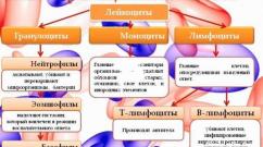
- production of red and white blood cells;
- accumulation of minerals, salts, water, organic compounds.
Bone has the ability to grow, change and regenerate. So, a small, newly born child has over 270 bones, and an adult has about 206. This is due to the fact that as they grow, many bones lose cartilage and fuse together.
Bone composition
The bones of the musculoskeletal system include the following elements:
- periosteum - outer film of connective tissue;
- endosteum - an internal connective tissue layer that forms the medullary canal inside the tubular bones;
- bone marrow is the soft tissue substance inside the bone;
- nerves and blood vessels;
- cartilage.
All bones are composed of organic (mainly collagen) and inorganic elements. The younger the body, the more organic compounds there are in the bones. In an adult, the collagen content in bones drops to 30%.
Bone structure
The structure of bone under a microscope looks like a set of concentric layers - plates inserted into each other, consisting of protein, mineral substance (hydroxyopatite) and collagen. This structural unit is called an osteon. The inner plate forms the so-called Haversian canal - a conductor for nerves and blood vessels. In total, an osteon can contain up to 20 similar plates, between which there are star-like bone cells. There are also insert plates between the osteons themselves. The lamellar structure, penetrated by the neurovascular Haversian canals, is characteristic of all bone surfaces, both external and internal, except for spongy bones. The presence of channels promotes the active participation of bones in mineral and bone metabolism and hematopoiesis (blood formation).
Cellular structure of bones
There are three types of cells in bones:
- Osteoblasts are immature young bone cells that synthesize the matrix - intercellular substance. They form on the surface of growing bones, as well as in places of bone damage. Over time, osteoblasts become cemented in the matrix and transform into osteocytes. These are the main participants in osteogenesis (bone synthesis).
- Osteocytes are mature, non-dividing, almost non-matrix-producing cells that communicate with each other through the channels of the cavities (lacunae) in which they are located. Tissue fluid circulates between the processes of osteocytes, its movement occurs due to the vibration of osteocytes. Osteocytes are living cells - thanks to them, metabolism is carried out and the mineral and organic balance in the bones is maintained.
- Osteoclasts are huge multinucleated cells that destroy old bone tissue. They, too, like osteoblasts, are important participants in bone formation. A balance must be maintained between osteoblasts and osteoclasts: if there are more osteoclasts than osteoblasts, osteoporosis begins in the bones.
Most bones develop from cartilaginous tissue, except for the bones of the skull, lower jaw and, presumably, the collarbone - they are formed from connective tissue.

Types of bones
The human musculoskeletal system is represented by bones of various types - long, flat, short, mixed, sesamoid.
- Long tubular bones have a rounded, hollow shape when cut. The middle elongated part of the bone (diaphysis) is filled inside with yellow bone marrow. At both ends of the tubular bone there is a head (epiphysis), covered on top with hyaline cartilage, and inside consisting of a spongy substance that contains red bone marrow. The growing part of the bone (metaphysis) is the area between the epiphysis and diaphysis. In children and adolescents, the metaphysis consists of cartilage, which is replaced by bone at the end of growth. The long tubular bones include the bones of the limbs, in particular the longest one, the femur.
- Flat bones are non-hollow, have a thin cut and consist of a spongy substance, covered on top with a compact smooth layer. The scapula, pelvic bones, and ribs have this structure.
- Short bones have a tubular or flattened structure, but there is no single cavity inside them. Cells with bone red marrow are separated by partitions. The short bones include the phalanges of the fingers, the carpus, the metacarpus, the tarsus, and the metatarsus.
- Mixed bones can combine elements of flat and short bones. Mixed bones include the vertebrae, occipital and temporal bones of the skull.
- Sesamoid bones are located deep in the tendon, at the point where it passes through the joint (knee, wrist, foot, etc.), they usually lie on the surface of another bone. Their task is to protect the tendon and strengthen the muscle by increasing the power arm.
All bones have irregularities in the form of protrusions, tubercles, depressions, and grooves. This is necessary for connecting bones and attaching muscle tendons.
A few notes about bone marrow
The bone marrow, unlike the brain and spinal marrow, has nothing to do with the central nervous system; it does not have neurons. This is a hematopoietic organ consisting of myeloid two-component tissue (stroma + hemal component).
In the growing bones of the skull and facial bones, mucous bone marrow is formed - a gelatinous consistency depleted of cells.
Main components of the human skeleton
The skeleton is the static basis of the human musculoskeletal system. The construction of the whole body begins with it. Skeletal anatomy must be adapted to each organ individually and to the entire set of vital systems, providing all the necessary functions of the musculoskeletal system.
Human skull
Let's start with the part that crowns the skeleton - the skull.
Humans are the highest mammals in the evolutionary chain, and this is reflected in our skull. The volume of the adult human brain is about 1500 cubic centimeters, so the brain portion of the human skull is relatively larger than that of animals. Relatively - this is in comparison with the front part. The human lifestyle inevitably led to the fact that in the process of evolution, people's brains grew and their jaws became smaller, because man, having learned to use tools, abandoned raw food.
The brain part of the skull consists of four unpaired and two paired bones fused together:
- unpaired - frontal, sphenoid, ethmoid and occipital;
- paired - two temporal and two parietal.
All the bones of the brain part of the adult skull are connected motionlessly, but in a newborn the sutures remain uncovered for a long time, connecting to each other through “fontanelles” - soft cartilaginous tissue - this is how nature took care of the growth of the skull.
In the occipital part of the skull there is an opening connecting the brain and the spinal cord; arteries supplying the brain with blood also pass through it. The skull is attached to the spine using an elliptical joint. Mobility is provided by the first two cervical vertebrae, called the atlas and epistrophy.
The facial part includes the following bones:
- paired bones: facial jaw, cheekbones, nasal bones, nasal cavity bones, palate;
- unpaired bones: lower jaw, hyoid bone, vomer.
The lower jaw is the only movable articular joint of the skull, and where there is a joint, there are diseases such as arthritis, dislocation, osteonecrosis, etc.
The spine is the basis of the ODS
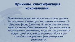
The spine is the axial rod of the human motor system. Unlike animals, it has a vertical position, which is also reflected in its structure: in profile, the spine in humans looks like the Latin letter S. These natural curves of the spine are designed to counteract the compressive forces to which the vertebrae are continuously exposed. They play the role of shock absorbers and balance the spine when dynamic load increases.

If there were no bends, our spine could break during a normal jump and it would be difficult to maintain balance.
In total, the spine has five vertebral sections and up to 34 vertebrae (maybe a couple less due to the different number of vertebrae in different people in the rudiment of the tail - the coccyx).
- the cervical spine has 7 vertebrae;
- chest - 12;
- lumbar and sacral - five vertebrae each;
- coccygeal - from 3 to 5.
Distribution of curves in the spine
The curvatures of the spine in adjacent sections are oppositely directed:
- cervical spine - the bend is directed forward, it is called lordosis.
- thoracic region - the bend is directed backwards, this is kyphosis. Exceeding the norm is called stooping.
- lumbar region - lordosis;
- sacral region - kyphosis.
Excessive bending in the lumbosacral region can lead to displacement of the vertebrae (spondylolisthesis), hernia, and destabilization of the spinal column.
The flexibility of the spinal column is also controlled by the vertebrae, which are semi-movably connected to each other using cartilaginous plates - intervertebral discs. Dystrophic changes in the discs lead to a catastrophic disease - osteochondrosis, from which all other orthopedic pathologies originate.
Let us now consider the remaining large elements included in the ODS.
The musculoskeletal system includes such important parts of the skeleton as the chest, shoulder girdle, upper and lower limbs, and pelvic girdle.
Rib cage
The chest is the repository of the organs of the chest cavity (heart, trachea, lungs). It is reinforced with a rib frame of 12 pairs of ribs:
- The first 7 pairs in front are semi-movably attached to the sternum;
- The 8th, 9th and 10th pairs of ribs are connected to each other by cartilage;
- the last two pairs are free.
At the back, all the ribs and vertebrae articulate, forming the costoarticular joint.
The thoracic region is inactive, so osteochondrosis in the chest is quite rare, but joint blockage, arthrosis, and intercostal neuralgia can be frequent sources of pain here.
Shoulder girdle
The shoulder girdle consists of two wedge-shaped shoulder blades and two curved clavicular bones, connecting in front to the sternum and behind to the shoulder blades. The upper limb is tied to the shoulder girdle. The shoulder joint is the loosest joint in the human body - this determines multidimensional free movement of the arm, but at the same time it threatens with problems such as shoulder dislocation, glenohumeral periarthritis, etc.

Upper limbs
Everyone seems to know what the upper limbs are made of, but anatomical terms do not always coincide with people’s definitions: many people call the collarbone the shoulder, and the upper arm the forearm. In fact, the hand consists of:
- from the humerus (the upper part of the arm that fits into the shoulder joint);
- the forearm, which includes two bones - the ulna and radius;
- carpal bone.
The hand has a lot of small bones:
- the wrist consists of eight bones, seven of which are arranged in two rows;
- metacarpus - made of 5 bones;
- fingers - from phalanges (two in the thumbs, three in the rest).
Such a terrible disease as rheumatoid arthritis begins precisely in the small wrist joints, so they can be a good indicator of this pathology.
Pelvic girdle
Located approximately in the middle of the body skeleton, the pelvic girdle plays an important role in distributing all loads on the spine (the center of gravity of the body is located just above it) and in balancing the spine. In addition, the pelvis protects important organs of the genitourinary system. Through the caudal foramen at the bottom, the hip and pelvis joint is attached to the spine.
The pelvic girdle consists of fused paired bones - the ilium, ischium and pubis. The hip joint (HJ) is made up of the acetabulum (the socket in the ilium) and the head of the femur.
Problems with the hip joint that lead to disability are coxarthrosis and hip dislocation. In addition, there are congenital anomalies associated with displacements and underdevelopment of the pelvic bones, leading to severe forms of scoliosis.
Lower limbs
The lower limbs include the femur and tibia (tibia and fibula) and feet, connected by the knee joints.
Foot composition:
- seven bones of the forearm, of which the calcaneus is the largest;
- five metacarpal bones;
- 14 phalanges of fingers (two in the big ones, three in all the others).
The knee joint, as well as the ankle, are the most loaded joints in the human body, so arthrosis, tendonitis, heel spurs, sprains and ligament tears make up the lion's share of problems with the lower extremities.
Muscle structure of the ODS
The musculoskeletal system also includes muscles: they are inextricably linked with the skeleton, without them it would simply collapse into a pile of bones. They are also not only a holding force, but also an active driving force.
Muscles consist of elastic tissue, microscopically represented by muscle cells - myocytes.
Muscle types
There are three types of muscles:
- skeletal or striated;
- smooth;
- cardiac.
The movement of absolutely all parts of our skeleton, including facial expressions, is carried out precisely by striated muscles. Skeletal muscles make up the majority of all muscles - there are more than 600 of them, and the total relative weight in the human body is about 40%. The smoothness and coordination of all movements is created due to the presence of agonist and antagonist muscles, which create two multidirectional efforts: the agonists perform the movement, the antagonists resist it.

The motor function of skeletal muscles is caused by their ability to contract in response to a signal from a nerve impulse coming from the central nervous system. The work of the muscles of this group is completely subject to the control of the human brain.
Striated muscles are 70-80% water, and the remaining 20% are proteins, glycogen, phosphoglycerides, cholesterol and other substances.
The most-most muscles of the body:
- The calf and chewing muscles are recognized as the strongest.
- The largest is the gluteal;
- The smallest are the ears;
- The longest muscle is the sartorius muscle, stretching from the ilium to the tibia.
Smooth muscle is a tissue that is part of all internal organs, skin and blood vessels. Spindle-shaped muscle cells make slow movements, not subject to human will and control - they are controlled only by the autonomic nervous system (ANS). Without smooth muscles, digestion, blood circulation, bladder function and other vital processes are impossible.
The cardiac muscle is included in a separate group, since it is striated, and at the same time it is not subordinate to human consciousness, but is subordinate only to the ANS. Also unique is the ability of the muscle to contract when removed from the chest cavity.
Muscle classification
There are a lot of muscles in the human body. They can be combined into separate groups according to their functions, the direction of the fibers, their relationship to the joints and their shape. Let's summarize the classification in a table:
| Classification type | Muscle names |
| By function: | Flexors, extensors, adductors, abductors, rotators, erectors, elevators, depressors, sphincters and dilators, synergists and antagonists |
| By fiber direction: | Rectus, transverse, teres, oblique (unipennate, bipennate, multipennate, semitendinous, semimembranosus) |
| In relation to joints: | One-piece, two-piece, multi-piece |
| By form: | Simple:
|
The human musculoskeletal system is a complex symbiosis of different systems: skeletal, muscular, nervous, and autonomic. It is inextricably linked with a person; any life process depends on it. It is designed simply beautifully, developing with us. There is nothing superfluous in it, so damage to a single part of it can destabilize the entire SDS and cause a number of subsequent diseases.
All organs of movement that ensure the movement of the body in space are combined into a single system. This includes bones, joints, muscles and ligaments. The human musculoskeletal system performs certain functions due to the peculiarities of the formation and structure of the organs of movement.
Importance of the musculoskeletal system
The human skeleton performs several vital functions:
- supporting;
- protective;
- provides movement;
- takes part in hematopoiesis.
Disorders of the musculoskeletal system cause pathological processes in the functioning of many body systems. Muscles attached to bones move them relative to each other, which ensures the movement of the body in space. The muscular apparatus has its own functional feature:
- surrounds the cavities of the human body, protecting them from mechanical damage;
- perform a supporting function, supporting the body in a certain position.
During the development of the human musculoskeletal system, the development of the central nervous system is stimulated. The development of muscles and nerve cells are mutually dependent processes. Knowing what functions of the musculoskeletal system are necessary for the normal functioning of the body, we can conclude that the skeleton is a vital structure of the body.
During the period of embryogenesis, when the body is practically not affected by any irritants, fetal movements cause irritation of muscle receptors. From them, impulses go to the central nervous system, stimulating the development of neurons. At the same time, the developing nervous system stimulates the growth and development of the muscular system.
Skeletal anatomy
Skeleton is a set of bones that perform supporting, motor and protective functions. The human musculoskeletal system has about 200 bones (depending on age), of which only 33-34 bones are unpaired. There are axial (chest, skull, spine) and accessory (free limbs) skeletons.
Bones are formed from a type of connective tissue. It consists of cells and a dense intercellular substance, which contains many mineral components and collagen, which provides elasticity.
The skeleton is a container for vital human organs: the brain is located in the skull, the spinal cord is located in the spinal canal, the chest provides protection to the esophagus, lungs, heart, main arterial and venous trunks, and the pelvis protects the organs of the genitourinary system from damage. Disorders of the musculoskeletal system can cause damage to internal organs, sometimes incompatible with life.
Bone structure
The bones contain a spongy and compact substance. Their ratio varies depending on the location and functions of a certain part of the musculoskeletal system.
The compact substance is localized in the diaphysis, which provides support and locomotor functions. Spongy substance is located in flat and short bones. The entire surface of the bone (with the exception of the articular surface) is covered with periosteum (periosteum).
Bone Formation
In ontogenesis, the formation of the musculoskeletal system goes through several stages - membranous, cartilaginous and bone. From the second week after conception, cartilaginous rudiments form in the mesenchyme of the membranous skeleton. By the 8th week, cartilage tissue is gradually replaced by bone tissue.
Replacement of cartilage tissue with bone tissue can take place in several ways:
- perichondrial ossification - the formation of bone tissue along the perimeter of the cartilage;
- periosteal ossification - production of young osteocytes by the formed periosteum;
- enchondral ossification - the formation of bone tissue within cartilage.
The process of bone tissue formation involves the growth of blood vessels and connective tissue from the periosteum into the cartilage (cartilage destruction occurs in these places). From some of the osteogenic cells, spongy bone subsequently develops.
During the period of intrauterine development of the fetus, ossification of the diaphyses of the tubular bones occurs (ossification points are called primary), then after birth, ossification of the epiphyses of the tubular bones occurs (secondary ossification points). Until the age of 16-24 years, a cartilaginous epiphyseal plate remains between the epiphyses and diaphyses.
Thanks to its presence, the organs of the musculoskeletal system are lengthened. After the bone is replaced and the diaphyses and epiphyses of the tubular bones fuse, human growth stops.
The structure of the spinal column
The spinal column is a series of overlapping vertebrae that are connected by the intervertebral discs, joints and ligaments that form the basis of the musculoskeletal system. The functions of the spine are not only support, but also protection, preventing mechanical damage to internal organs and the spinal cord passing in the spinal canal.

There are five sections of the spine - coccygeal, sacral, lumbar, thoracic and cervical. Each section has a certain degree of mobility; only the sacral spine is completely immobile.
The movement of the spine or its parts is ensured with the help of skeletal muscles. The correct development of the musculoskeletal system in the neonatal period provides the necessary support for internal organs and systems and their protection.
Structure of the chest
The rib cage is an osteochondral formation consisting of the sternum, ribs and 12 thoracic vertebrae. The shape of the chest resembles an irregular truncated cone. The chest has 4 walls:
- anterior - formed by the sternum and cartilage of the ribs;
- posterior - formed by the vertebrae of the thoracic spine and the posterior ends of the ribs;
- 2 lateral - formed directly by the ribs.

In addition, there are two openings of the chest - the upper and lower apertures. The organs of the respiratory and digestive system (esophagus, trachea, nerves and blood vessels) pass through the upper opening. The lower aperture is closed by a diaphragm, in which there are openings for the passage of large arterial and venous trunks (aorta, inferior vena cava) and the esophagus.
Structure of the skull
The skull is one of the main structures that forms the musculoskeletal system. The functions of the skull are to protect the brain, sensory organs and support the initial parts of the respiratory and digestive systems. It consists of paired and unpaired bones and is divided into the brain and facial sections.
The facial section of the skull consists of:
- from the maxillary and mandibular bones;
- two nasal bones;
The brain section of the skull includes:
- paired temporal bone;
- paired sphenoid bone;
- steam room;
- occipital bone.

The brain section performs a protective function for the brain and is its container. The facial region provides support for the initial portion of the respiratory and digestive systems and sensory organs.
Musculoskeletal system: functions and structure of the limbs
In the process of evolution, the skeleton of the limbs acquired extensive mobility due to the articulation of the bones (especially the radial and carpal joints). The thoracic and pelvic girdles are distinguished.
The upper girdle (pectoral) includes the scapula and two clavicle bones, and the lower (pelvic) is formed by the paired pelvic bone. The following sections are distinguished in the free part of the upper limb:
- proximal - represented by the humerus;
- middle - represented by the ulna and radius bones;
- distal - includes the carpal bones, metacarpal bones and finger bones.

The free part of the lower limb consists of the following sections:
- proximal - represented by the femur;
- middle - includes the tibia and fibula;
- distal - tarsal bones, metatarsal bones and finger bones.

The skeleton of the limbs provides the possibility of a wide range of actions and is necessary for normal work activity, which is provided by the musculoskeletal system. The functions of the skeleton of free limbs are difficult to overestimate, since with their help a person performs almost all actions.
The structure of the muscular system
Skeletal muscles are attached to bones and, when contracted, provide movement of the body or its individual parts in space. Skeletal muscles are based on striated muscle fibers. In addition to supporting and motor functions, muscles provide the function of breathing, swallowing, chewing, and take part in facial expressions, heat production and speech articulation.
The main properties of skeletal muscles are:
- excitability - the activity of muscle fibers is carried out under the influence of nerve impulses;
- conductivity - from the nerve endings to the central nervous system there is a rapid conduction of the impulse;
- contractility - as a result of the movement of a nerve impulse, contractility of the skeletal muscle occurs.

A muscle consists of tendinous ends (tendons that attach the muscle to the bone) and a belly (consisting of striated muscle fibers). The coordinated work of the musculoskeletal system is carried out by the correct functioning of the muscles and the necessary nervous regulation of muscle fibers.