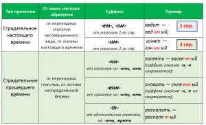
Physiology and age-related features of the blood system. Age-related blood characteristics in children and adolescents
As you know, with age a person gets older. His heart function deteriorates, visual acuity and hearing decrease. Memory fails more and more often. The joints begin to ache. The skin wrinkles and becomes decrepit. However, not only internal organs and skin undergo aging, but also the blood fluid that flows in every person. Age-related features of the blood system are peculiar. You can't say enough about them in a few words. This reduces the normal composition of the blood: leukocytes, erythrocytes, platelets, which affects the immune system, cell nutrition, blood clotting and other structures of the body. Age and other characteristics of the blood system lead to a number of complex diseases.
The normal blood composition cannot be the same in newborns, adolescents and adults. Its indicators change over time, and depending on age, the required values are formed. A visual table shows the current sequence well.
In mature men and women weighing 65-75 kilograms, the blood level will be five to six liters. Aging also affects the percentage of the main elements of blood fluid. In adults, healthy people of both sexes, the norm of blood cells (erythrocytes, leukocytes, platelets) is: 41-43 percent in women and 44-46 percent in men. The entire remaining volume of the level is plasma. The indicator of the volume of elements to plasma is called the hematocrit number.
Over the course of life, the numerical value may change. For example, in a child, immediately after birth it is 54%. This is due to the high number of red blood cells. By the beginning of the second week of life, the norm decreases and reaches 52 percent. By the beginning of the second month 42%. In the annual period, the ratio of formed elements is indicated by the number 35%. By the beginning of the sixth year of life - 37%, and by the age of fifteen it can reach 39 percent. The normal level of adult indicators of 40-45% is formed by approximately 15-year-old adolescents.
Age-related features of the blood system also affect formed substances. Thus, the indicators of red blood cells in adult men and women are not the same. For the weaker sex, the normal level is listed as 3.7-4.7 million per 1 mm 3. The stronger sex has 4.0-5.1 million per 1 mm 3.
In newborns, the number of red cells ranges from 4.3-7.6 million per 1 mm 3 of blood fluid. In a six-month-old child, red blood cells drop to 3.5-4.8 million per 1 mm 3. In one-year-old children, 3.6-4.9 million per 1 mm 3. In adolescence, closer to 15 years, their normal level reaches values similar to those of adults, relative to the gender of the child.
About leukocytes and red blood cells
The same can be said about the hemoglobin content. In an adult, it can be 16.7 g per 100 ml of blood. For women the norm is 70-80 percent, for men 80-100%. These indicators depend on the number of red blood cells. In a general sense, hemoglobin levels are affected by many conditions. So, in newborns it can be in the range of 110-140 percent. By six months it decreases to 70-80%. By the age of four, its norm increases to 85%. In six to seven year old children it drops slightly, and from the age of eight we can say that hemoglobin levels begin to rise. In adolescence they can be in the range of 70-90%.

We can say that age also imposes restrictions on the development of leukocytes. If we take the internal mobile environment of an adult as a basis, then one μl can contain from 4000 to 9000 leukocytes. Newborns contain up to 20 thousand leukocytes per cubic millimeter of blood. Sometimes it increases to 30 thousand in 1 mm 3. Then we can talk about limitation and declining dynamics. By the second week of a baby’s life, their number is 10-12 thousand.
Gradually, the number of white cells decreases and by adolescence their value can be the same as in adults, taking into account gender. Also, in newborns, blood clotting is slow, but starting from the 3rd day of a baby’s life, this process accelerates and reaches the values of an adult. For preschoolers and schoolchildren, the time interval for blood fluid clotting is individual. On average, the formation of a platelet plug occurs after 1-2 minutes and ends after 4 minutes.
From birth to adulthood
Age-related features of blood vessels also deserve attention. We can say that until the moment a child becomes an adult, his vascular structure is gradually built up:
- arteries thicken;
- the length of the vessels increases;
- a rounded shape of the blood channels is formed.
In both sexes, the right coronary artery is smaller in diameter than the left coronary artery. But the difference is especially noticeable in infants and adolescents. The carotid artery in diameter in adults is nine to fourteen millimeters. Babies have six millimeters. In children under ten years of age, of all the cerebral arteries, the largest is the middle one. The main arteries develop faster than their branches. In children from one to five years of age, the ulnar artery grows faster than the radial artery, but then the radial artery will prevail.

The length of the arteries and its development depend on the growth of the child. Those bloodstreams that supply the brain develop quite actively, especially at an early stage of life. The leader in increasing length can be considered the anterior cerebral artery. But other arteries involved in the process of blood flow, especially the upper ones, do not lag behind. lower limbs, as well as organs. In infants, the inferior mesenteric artery extends six centimeters. In a mature body - by 17 cm. Along with this, the radius of curvature of the arcs also changes. In children and early adolescents, the aortic arch is significantly larger relative to the radius of curvature. In adults it is less.
Arches, vertebrae, canals
- In the smallest children, it dominates the level of the first thoracic vertebra.
- On the horizontal of the 2nd vertebra, at seventeen to twenty years old.
- Between 25 and 30 years of age, the aortic arch moves to the level of the third vertebra.
- Closer to 45 years, it decreases to the 4th thoracic vertebra.
- For those over fifty years of age and older, it is located between the 4th and 5th vertebrae.
The anatomy of the arteries is gradually changing. As we grow older, the radial and ulnar arteries shift relative to the midline of the forearm in a lateral manner. By the age of 10, these vessels occupy the same position as in an adult body.

The anatomical structure of the palmar arterial arches is also formed. In children and infants, the superficial arch lies closer to the middle of the 2nd and 3rd metacarpals. Next it moves to the level of the middle part of the 3rd metacarpal bone. The branching of arteries also changes with age. From the moment of birth, the toddler has a loose branching pattern. Not immediately, the main appearance of the arteries is structured and after ten years of age does not change. Intraorgan vessels also gradually increase in size. Intensively changing:
- diameter;
- length;
- number per unit volume.
These changes are active between eight and twelve life cycles. Microcirculation channels located in organs increase as the organs themselves develop.
The diameter of the veins of the systemic circulation increases gradually . Over the years, the area of the body increases, as well as the cross-sectional length. At a young age, the superior vena cava is short due to the high position of the heart muscle. In one-year-olds, boys and girls, its length and area increase throughout life cycle don't change. Only in old age is an expansion of the diameter observed. The other vena cava is the inferior one, which is short and wide in newborns.
During adulthood, its diameter increases faster than that of the superior vena cava. In newborns, its formation occurs at the 3-4 vertebrae. Further, the level decreases and in adolescence approaches the 4-5 vertebrae. As it forms, the angle of inclination also changes. In newborns it can be 45-75 degrees, in adults between 70 and 100 degrees. In general, age-related features of blood vessels are observed from the day of birth, before puberty and in old age.
Amount of blood. The amount of blood in an adult is on average 7% of body weight, in newborns - from 10 to 20% of body weight, in infants - from 9 to 13%, in children from 6 to 16 years old - 7%. The younger the child, the higher his metabolism and the greater the amount of blood per 1 kg of body weight. Newborns have 150 cubic meters per 1 kg of body weight. cm of blood, in infants - 110 cubic meters. cm, for children from 7 to 12 years old - 70 cubic meters. cm, from 15 years old - 65 cubic meters. cm. The amount of blood in boys and men is relatively greater than in girls and women. At rest, approximately 40-45% of the blood circulates in the blood vessels, and the rest is in the depot (capillaries of the liver, spleen and subcutaneous tissue). Blood from the depot enters the general bloodstream when body temperature rises, muscle work, rise to altitude, and blood loss. Rapid loss of circulating blood is life-threatening. For example, with arterial bleeding and loss of 1/3-1/2 of the total amount of blood, death occurs due to a sharp drop in blood pressure.
Blood plasma. Plasma is the liquid part of the blood after all the formed elements have been separated. In adults it accounts for 55-60% of the total blood volume, in newborns - less than 50% due to the large volume of red blood cells. The blood plasma of an adult contains 90-91% water, 6.6-8.2% proteins, of which 4-4.5% albumin, 2.8-3.1% globulin and 0.1-0.4% fibrinogen; the rest of the plasma consists of minerals, sugar, metabolic products, enzymes, and hormones. The protein content in the plasma of newborns is 5.5-6.5%, in children under 7 years old – 6-7%.
With age, the amount of albumin decreases and globulin increases, the total protein content approaches the level of adults by 3-4 years. Gamma globulins reach the adult norm by 3 years, alpha and beta globulins by 7 years. The blood levels of proteolytic enzymes increase after birth and reach adult levels by the 30th day of life.
Blood minerals include table salt (NaCl), 0.85-0.9%, potassium chloride (KC1), calcium chloride (CaC12) and bicarbonates (NaHCO3), 0.02% each, etc. In newborns, the amount of sodium less than in adults, and reaches normal by 7-8 years. From 6 to 18 years of age, sodium content ranges from 170 to 220 mg%. The amount of potassium, on the contrary, is highest in newborns, lowest at 4-6 years of age and reaches the adult norm by 13-19 years.
Boys aged 7-16 years have 1.3 times more inorganic phosphorus than adults; organic phosphorus is 1.5 times more than inorganic phosphorus, but less than in adults.
The amount of glucose in the blood of an adult on an empty stomach is 0.1–0.12%. The amount of blood sugar in children (mg%) on an empty stomach: in newborns - 45-70; for children 7-11 years old - 70-80; 12–14 years old - 90-120. The change in blood sugar levels in children aged 7-8 years is significantly greater than in children aged 17-18 years. Significant fluctuations in blood sugar levels occur during puberty. With intense muscular work, blood sugar levels decrease.
The viscosity of the blood of an adult is 4-5, of a newborn - 10-11, of a child in the first month of life - 6, then a gradual decrease in viscosity is observed.
Blood, lymph and tissue fluid are the internal environment of the body in which the vital activity of cells, tissues and organs takes place. The internal environment of a person maintains a relative constancy of its composition, which ensures the stability of all functions of the body and is the result of reflex and neurohumoral self-regulation. Blood, circulating in blood vessels, performs a number of vital functions: transport (transports oxygen, nutrients, hormones, enzymes, and also delivers residual metabolic products to the excretory organs), regulatory (maintains a relative constancy of body temperature), protective (blood cells provide immune responses).
Amount of blood. Deposited and circulating blood. The amount of blood in an adult is on average 7% of body weight, in newborns - from 10 to 20% of body weight, in infants - from 9 to 13%, in children from 6 to 16 years old - 7%. The younger the child, the higher his metabolism and the greater the amount of blood per 1 kg of body weight. Newborns have 150 cubic meters per 1 kg of body weight. cm of blood, in infants - 110 cubic meters. cm, for children from 7 to 12 years old - 70 cubic meters. cm, from 15 years old - 65 cubic meters. cm. The amount of blood in boys and men is relatively greater than in girls and women. At rest, approximately 40–45% of the blood circulates in the blood vessels, and the rest is in the depot (capillaries of the liver, spleen and subcutaneous tissue). Blood from the depot enters the general bloodstream when body temperature rises, muscle work, rise to altitude, and blood loss. Rapid loss of circulating blood is life-threatening. For example, with arterial bleeding and loss of 1/3-1/2 of the total amount of blood, death occurs due to a sharp drop in blood pressure.
Blood plasma. Plasma is the liquid part of the blood after all the formed elements have been separated. In adults it accounts for 55–60% of the total blood volume, in newborns it is less than 50% due to the large volume of red blood cells. The blood plasma of an adult contains 90–91% water, 6.6–8.2% proteins, of which 4–4.5% albumin, 2.8–3.1% globulin and 0.1–0.4% fibrinogen; the rest of the plasma consists of minerals, sugar, metabolic products, enzymes, and hormones. The protein content in the plasma of newborns is 5.5–6.5%, in children under 7 years of age – 6–7%.
With age, the amount of albumin decreases and globulin increases; the total protein content approaches the level of adults by 3–4 years. Gamma globulins reach the adult norm by 3 years, alpha and beta globulins by 7 years. The blood levels of proteolytic enzymes increase after birth and reach adult levels by the 30th day of life.
Blood minerals include table salt (NaCl), 0.85-0.9%, potassium chloride (KC1), calcium chloride (CaC12) and bicarbonates (NaHCO3), 0.02% each, etc. In newborns, the amount of sodium less than in adults, and reaches normal by 7–8 years. From 6 to 18 years of age, sodium content ranges from 170 to 220 mg%. The amount of potassium, on the contrary, is highest in newborns, lowest at 4–6 years of age and reaches the adult norm by 13–19 years.
Boys aged 7-16 years have 1.3 times more inorganic phosphorus than adults; organic phosphorus is 1.5 times more than inorganic phosphorus, but less than in adults.
The amount of glucose in the blood of an adult on an empty stomach is 0.1–0.12%. The amount of blood sugar in children (mg%) on an empty stomach: in newborns – 45–70; for children 7-11 years old – 70–80; 12–14 years old – 90-120. The change in blood sugar levels in children aged 7–8 years is significantly greater than in children aged 17–18 years. Significant fluctuations in blood sugar levels occur during puberty. With intense muscular work, blood sugar levels decrease.
In addition, blood plasma contains various nitrogenous substances, amounting to 20–40 mg per 100 cubic meters. cm blood; 0.5–1.0% fat and fat-like substances.
The viscosity of the blood of an adult is 4–5, of a newborn – 10–11, of a child in the first month of life – 6, then a gradual decrease in viscosity is observed. The active blood reaction, depending on the concentration of hydrogen and hydroxyl ions, is slightly alkaline. The average blood pH is 7.35. When acids formed during metabolism enter the blood, they are neutralized by a reserve of alkalis. Some acids are removed from the body, for example carbon dioxide is converted to carbon dioxide and water vapor exhaled during increased ventilation. When there is excessive accumulation of alkaline ions in the body, for example during a vegetarian diet, they are neutralized by carbonic acid, which is retained when ventilation of the lungs decreases.
7.2. Formed elements of blood
The formed elements of blood include red blood cells, leukocytes and platelets. Erythrocytes are non-nucleated red blood cells. They have a biconcave shape, which increases their surface by approximately 1.5 times. The number of red blood cells in 1 cubic meter. mm of blood is equal to: in men – 5–5.5 million; in women - 4–5.5 million. In newborns on the first day of life, their number reaches 6 million, then a decrease occurs to the adult norm. At 7–9 years old, the number of red blood cells is 5–6 million. The greatest fluctuations in the number of red blood cells are observed during puberty.
In the red blood cells of an adult, hemoglobin makes up about 32% of the weight of the formed elements and on average 14% of the weight of whole blood (14 g per 100 g of blood). This amount of hemoglobin is equal to 100%. The hemoglobin content in the red blood cells of newborns reaches 14.5% of the adult norm, which is 17–25 g of hemoglobin per 100 g of blood. In the first two years, the amount of hemoglobin drops to 80–90%, and then rises again to normal. The relative content of hemoglobin increases with age and by 14–15 years it reaches the adult norm. It is equal (in grams per 1 kg of body weight):
at 7–9 years old – 7.5;
10–11 years old – 7.4;
12–13 years old – 8.4;
14–15 years old – 10.4.
Hemoglobin is species specific. If in a newborn it absorbs more oxygen than in an adult (and from the age of 2 this ability of hemoglobin is maximum), then from the age of 3 hemoglobin absorbs oxygen in the same way as in adults. The significant content of red blood cells and hemoglobin, as well as the greater ability of hemoglobin to absorb oxygen in children under 1 year of age, provide them with a more intense metabolism.
With age, the amount of oxygen in arterial and venous blood increases. 0 but equals (in cubic cm per minute): in children 5–6 years old in arterial blood - 400, in venous blood - 260; in adolescents 14–15 years old – 660 and 435, respectively; in adults – 800 and 540, respectively. The oxygen content in arterial blood (in cubic cm per 1 kg of weight per minute) is equal to: in children 5–6 years old – 20; in adolescents 14–15 years old – 13; in adults - 11. This phenomenon in preschool children is explained by the relatively large amount of blood and blood flow, significantly exceeding the blood flow of adults.
In addition to transporting oxygen, red blood cells are involved in enzymatic processes, maintaining an active blood reaction and in the exchange of water and salts. During the day, from 300 to 2000 cubic meters pass through red blood cells. dm of water.
In the process of settling whole blood, to which substances that prevent blood clotting have been added, red blood cells gradually settle. The rate of erythrocyte sedimentation reaction (ESR) in men is 3–9 mm, in women – 7–12 mm per hour. S0E depends on the amount of proteins in the blood plasma and on the ratio of globulins to albumins. Since a newborn’s plasma contains about 6% proteins and the ratio of globulins to albumins is also less than in adults, their ESR is about 2 mm, in infants – 4–8 mm, and in older children – 4–8 mm per hour After an educational load, in most children 7-11 years old, normal (up to 12 mm per hour) and slow ESR accelerate, and accelerated ESR slows down.
Hemolysis. Red blood cells are able to survive only in physiological solutions, in which the concentration of minerals, especially table salt, is the same as in blood plasma. In solutions where the sodium content is less or more than in the blood plasma, as well as under the influence of other factors, red blood cells are destroyed. The destruction of red blood cells is called hemolysis.
The ability of red blood cells to resist hemolysis is called resistance. With age, the resistance of erythrocytes decreases significantly: the erythrocytes of newborns have the greatest resistance; by the age of 10 it decreases by about 1.5 times.
In a healthy body, there is a constant process of destruction of red blood cells, which is carried out under the influence of special substances - hemolysins, produced in the liver. Red blood cells live for 14 days in a newborn, and no more than 100–150 days in an adult. Hemolysis occurs in the spleen and liver. Simultaneously with hemolysis, new red blood cells are formed, so the number of red blood cells is maintained at a relatively constant level.
Blood groups. Depending on the content of two types of adhesive substances (agglutinogens A and B) in erythrocytes, and two types of agglutinins (alpha and beta) in plasma, four blood groups are distinguished. When transfusing blood, it is necessary to avoid matching A with alpha and B with beta, because agglutination occurs, leading to blockage of blood vessels and preceding hemolysis in the recipient, and therefore leading to his death.
Red blood cells of the first group (0) are not glued together by the plasma of other groups, which allows them to be administered to all people. People with the first blood group are called universal donors. Plasma of the fourth group (AB) does not glue red blood cells of other groups, so people with this blood group are universal recipients. Blood of the second group (A) can be transfused only to groups A and AB, blood of group B - only B and AB. Blood type is determined genetically.
In addition, in the practice of blood transfusion, the agglutinogen Rh factor (Rh) is of particular importance. The red blood cells of 85% of people contain the Rh factor (Rh positive), while the red blood cells of 15% of people do not contain it (Rh negative).
Leukocytes. These are colorless nucleated blood cells. In an adult, 1 cu. mm of blood contains 6–8 thousand leukocytes. Based on the shape of the cell and nucleus, leukocytes are divided into: neutrophils; basophils; eosinophils; lymphocytes; monocytes.
Unlike adults, newborns have 1 cubic meter. mm of blood contains 10–30 thousand leukocytes. The most large number leukocyte count is observed in children aged 2–3 months, and then it gradually decreases in waves and reaches the level of adults by 10–11 years.
In children under 9-10 years of age, the relative content of neutrophils is significantly lower than in adults, and the number of lymphocytes increases sharply until 14-15 years of age. Up to 4 years, the absolute number of lymphocytes exceeds the number of neutrophils by approximately 1.5–2 times; from 4 to 6 years, the number of neutrophils and lymphocytes is first compared, and then neutrophils begin to predominate over lymphocytes, and from the age of 15 their ratio approaches adult norms. Leukocytes live up to 12–15 days.
Unlike erythrocytes, the content of leukocytes fluctuates greatly. A distinction is made between an increase in the total number of leukocytes (leukocytosis) and a decrease (leukopenia). Leukocytosis is observed in healthy people during muscular work, in the first 2–3 hours after eating and in pregnant women. A person lying down has twice as much leukocytosis as a person standing. Leukopenia occurs when exposed to ionizing radiation. Some diseases change the relative abundance of different forms of white blood cells.
Platelets. These are the smallest nuclear-free plates of protoplasm. In adults, 1 cu. mm of blood contains 200–100 thousand platelets, in children under 1 year of age – 160–330 thousand; from 3 to 4 years – 350–370 thousand. Platelets live 4–5 and no more than 8–9 days. The dry residue of platelets contains 16–19% lipids (mainly phosphatides), proteolytic enzymes, serotonin, blood clotting factors and retractin. An increase in the number of platelets is called thrombocytosis, a decrease is called thrombopenia.
7.3. Circulation
Blood is able to perform vital functions only when it is in constant motion. The movement of blood in the body, its circulation constitute the essence of blood circulation.
The circulatory system maintains the constancy of the internal environment of the body. Thanks to blood circulation, oxygen, nutrients, salts, hormones, water are supplied to all organs and tissues and metabolic products are removed from the body. Due to the low thermal conductivity of tissues, heat transfer from the organs of the human body (liver, muscles, etc.) to the skin and into environment carried out mainly due to blood circulation. The activity of all organs and the body as a whole is closely related to the function of the circulatory system.
Large and small circles of blood circulation. Blood circulation is ensured by the activity of the heart and blood vessels. Vascular system consists of two circles of blood circulation: large and small.
The systemic circulation begins from the left ventricle of the heart, from where blood enters the aorta. From the aorta, the path of arterial blood continues through the arteries, which branch as they move away from the heart, and the smallest of them break up into capillaries, which permeate the entire body in a dense network. Through the thin walls of the capillaries, the blood releases nutrients and oxygen into the tissue fluid. In this case, the waste products of the cells enter the blood from the tissue fluid. From the capillaries, blood flows into small veins, which, merging, form larger veins and flow into the superior and inferior vena cava. The superior and inferior vena cava bring venous blood to the right atrium, where the systemic circulation ends.
The pulmonary circulation begins from the right ventricle of the heart by the pulmonary artery. Venous blood is carried through the pulmonary artery to the capillaries of the lungs. In the lungs, gases are exchanged between the venous blood of the capillaries and the air in the alveoli of the lungs. From the lungs, through four pulmonary veins, arterial blood returns to the left atrium, where the pulmonary circulation ends. From the left atrium, blood enters the left ventricle, where the systemic circulation begins.
7.4. Heart: structure and age-related changes
The heart is hollow muscular organ, divided into four chambers: two atria and two ventricles. The left and right sides of the heart are separated by a solid septum. Blood from the atria enters the ventricles through openings in the septum between the atria and ventricles. The holes are equipped with valves that open only towards the ventricles. Valves are formed by closing flaps and are therefore called leaflet valves. The left side of the heart has a bicuspid valve, and the right side has a tricuspid valve.
The semilunar valves are located at the exit of the aorta from the left ventricle and the pulmonary artery from the right ventricle. Semilunar valves allow blood to pass from the ventricles into the aorta and pulmonary artery and prevent the reverse flow of blood from the vessels into the ventricles.
Heart valves allow blood to flow in only one direction: from the atria to the ventricles and from the ventricles to the arteries.
The mass of the human heart ranges from 250 to 360 g.
The widened upper part of the heart is called the base, and the narrowed lower part is called the apex. The heart lies obliquely behind the sternum. Its base is directed back, up and to the right, and its top is directed down, forward and left. The apex of the heart is adjacent to the anterior chest wall in the area at the left intercostal space; here, at the moment of contraction of the ventricles, a cardiac impulse is felt.
The bulk of the heart wall is made up of a powerful muscle - the myocardium, consisting of a special kind of striated muscle tissue. The thickness of the myocardium varies in different parts of the heart. It is thinnest in the atria (2–3 mm). The left ventricle has the most powerful muscle wall: it is 2.5 times thicker than in the right ventricle.
Typical and atypical muscles of the heart. The bulk of the cardiac muscle is represented by fibers typical of the heart, which ensure contraction of the heart’s parts. Their main function is contractility. This is the typical working muscle of the heart. In addition to it, the cardiac muscle contains atypical fibers, the activity of which is associated with the occurrence of excitation in the heart and the conduction of excitation from the atria to the ventricles.
Atypical muscle fibers differ from contractile fibers both in structure and physiological properties. They have less pronounced transverse striations, but they have the ability to be easily excited and are more resistant to harmful influences. Due to the ability of atypical muscle fibers to conduct the resulting excitation through the heart, it is called the conduction system of the heart.
Atypical muscles occupy a very small part of the heart in volume. Clusters of atypical muscle cells are called nodes. One of these nodes is located in the right atrium, near the confluence (sinus) of the superior vena cava. This is the sinoatrial node. Here, in the heart of a healthy person, excitation impulses arise that determine the rhythm of heart contractions. The second node is located on the border between the right atrium and the ventricles in the septum of the heart, it is called the atrioventricular, or atrioventricular, node. In this area of the heart, excitation spreads from the atria to the ventricles.
From the atrioventricular node, excitation is directed along the atrioventricular bundle (bundle of His) of fibers of the conduction system, which is located in the septum between the ventricles. The trunk of the atrioventricular bundle is divided into two legs, one of them goes to the right ventricle, the other to the left.
Excitation from the atypical muscles is transmitted to the fibers of the contractile muscles of the heart with the help of fibers belonging to the atypical muscles.
Age-related changes in the heart. After birth, a child’s heart not only grows, but also undergoes morphological processes (shape and proportions change). The newborn's heart occupies a transverse position and has an almost spherical shape. The relatively large liver makes the vault of the diaphragm high, so the position of the heart in a newborn is higher (it is located at the level of the fourth left intercostal space). By the end of the first year of life, under the influence of sitting and standing and due to the lowering of the diaphragm, the heart takes an oblique position. By 2–3 years, the apex of the heart reaches the fifth rib. In ten-year-old children, the boundaries of the heart become almost the same as in adults.
During the first year of life, the growth of the atria outstrips the growth of the ventricles, then they grow almost equally, and after 10 years the growth of the ventricles begins to outstrip the growth of the atria.
Children's hearts are relatively larger than adults'. Its mass is approximately 0.63-0.80% of body weight, in an adult it is 0.48-0.52%. The heart grows most rapidly in the first year of life: by 8 months the heart’s mass doubles, by 3 years it triples, by 5 years it quadruples, and at 16 years – by 11 times.
The heart mass in boys in the first years of life is greater than in girls. At the age of 12–13 years, a period of increased growth of the heart begins in girls, and its mass becomes larger than in boys. By the age of 16, girls’ hearts again begin to lag behind boys’ hearts in mass.
Cardiac cycle. The heart contracts rhythmically: contractions of the heart parts (systole) alternate with their relaxation (diastole). The period covering one contraction and one relaxation of the heart is called the cardiac cycle. In a state of relative rest, the adult heart beats approximately 75 times per minute. This means that the entire cycle lasts about 0.8 s.
Each cardiac cycle consists of three phases:
1) atrial systole (lasts 0.1 s);
2) ventricular systole (lasts 0.3 s);
3) general pause (0.4 s).
During heavy physical activity, the heart contracts more than 75 times per minute, and the duration of the total pause decreases.
Lecture No. 8
Age-related features of blood and circulation
Key words: homeostasis, blood circulation, heart valves
Plan
- Composition and functions of blood.
Circulatory system
- Composition and functions of blood.
The constancy of the composition of blood plasma is ensured by the regulatory influence of the nervous system, as well as hormones. The respiratory and excretory organs play a role in maintaining this constancy.
Blood functions:
1. Respiratory function:
- transfer of oxygen from the lungs to the tissues and removal of carbon dioxide from the tissues to the lungs;
- oxygen transfer is carried out by hemoglobin contained in red blood cells (oxyhemoglobin is formed with oxygen);
- carbon dioxide is transported by plasma.
2.Nutritional function:
- transfer of nutrients: glucose, amino acids, fats, salts, vitamins, etc. from the digestive organs to the tissues.
- transfer of unnecessary metabolic products (urea, uric acid, etc.), as well as excess amounts of water and salts to the excretory organs.
5. Participation of blood in the regulation of body temperature. Blood plasma mainly consists of water, which has high thermal conductivity.
6. Regulatory role of blood: transport of hormones, enzymes and other biologically active substances throughout the body.
Blood composition.
Plasma and formed elements (blood cells). The formed elements are erythrocytes, leukocytes and platelets. In an adult, formed elements make up 46% of the blood volume, and plasma - about 54%. All blood cells live for a certain time and are destroyed. In hematopoietic tissues (bone marrow, lymph nodes, spleen), new cells are formed.
Amount of blood.
In children, the amount of blood is relatively larger than in adults; this is biologically determined. Blood, through its functions, ensures the flow of metabolism in tissues.
During different periods of growth and development, the intensity of metabolic processes in children is higher, and therefore the blood content in the body is higher.
Blood plasma.
Plasma is the liquid part of blood. Consists of 90% water and 10% dry matter (> proteins and salts). The osmotic pressure of the blood depends on the content of minerals.
Age-related features of blood circulation.
Fetal blood circulation has its own characteristics due to the fact that before birth, oxygen enters the fetal body through the placenta and umbilical vein. The umbilical vein branches into two vessels, one supplies the liver, the other connects to the inferior vena cava. In the inferior vena cava, oxygen-rich blood mixes with blood that has passed through the liver and contains metabolic products. Through the inferior vena cava, mixed blood enters the right atrium, then into the right ventricle and is pushed into the pulmonary artery: a smaller part of the blood flows into the lungs, and most of it through the ductus Botallus enters the aorta. The ductus botallus connects the pulmonary artery to the aorta. As a result of the connection of the pulmonary artery and the aorta, both ventricles pump blood into the systemic circulation. Blood with metabolic products returns to the maternal body through the umbilical arteries and placenta.
So, the main features of the fetal blood circulation: 1) circulation of mixed blood in the fetal body; 2) connection of the fetus through the placenta with the mother’s circulatory system; 3) the presence of the Botallov duct connecting the pulmonary artery to the aorta.
Circulatory system.
The heart is a muscular organ divided into 4 cavities - the right and left atrium, the right and left ventricle. The atrium is separated by the interatrial septum and the ventricles by the interventricular septum.
The average weight of the heart in women is 250 g, in men - 300 g. Through the atrioventricular openings equipped with valves, blood from the atria enters the ventricles. Valves are formed by leaflets and are called leaflet valves. Between the left atrium and the left ventricle is bivalve/mitral/ valve, on the right between the atrium and ventricle tricuspid. The valves allow blood to flow in only one direction: from the atria to the ventricles, and from the ventricles to the arteries. Arteries originate from the ventricles of the heart (an artery is a vessel that carries blood from the heart):
- from the left ventricle – the largest artery /aorta/,
from the right ventricle - pulmonary artery;
- At the border between the ventricles and arteries there are semilunar valves.
. Through small veins (venules) and large veins, blood returns to the heart. The superior and inferior vena cava approach the right atrium, and the four pulmonary veins approach the left atrium. Blood moves through two circles of blood circulation: large and small . Systemic circulation: from the left ventricle to all organs and tissues and ends in the right atrium. Small circle: from the right ventricle through the lungs / oxygen enrichment / to the left atrium.
Age-related features of the heart.
In children, the mass of the heart and the total lumen of blood vessels are larger than in adults, which facilitates blood circulation processes. In a newborn, the heart is spherical and located much higher than in an adult. The heart rate in children is higher than in adults, which is associated with the predominance of the tone of the sympathetic centers in children. Blood pressure in children is lower than in adults, and the flow rate is higher.
Properties of the heart muscle.
The heart wall consists of 3 layers: the outer or epicardium, the middle - the myocardium, and the inner - the endocardium. The bulk of the heart wall is a powerful muscle - the myocardium, consisting of striated muscle tissue. The left ventricle has the most powerful muscle wall.
The main function of the heart muscle is contractility. The heart muscle contains special atypical fibers in which excitation occurs. Atypical tissue consists of:
1) sinoatrial node (Kisa-Flaca node), located on the posterior wall of the right atrium;
2) atrioventricular node (Aschoff-Tavara node), located near the septum between the atria and ventricles;
3) atrioventricular bundle (bundle of His) extending from the natural ventricular node along the same trunk.
The bundle of His, passing through the septum between the atria and ventricles, divides into 2 legs leading to the ventricles. Nerve fibers from the vagus and sympathetic nerves approach the nodes of atypical tissue.
Cardiac cycle.
The cardiac cycle is a set of electrical, mechanical, and biochemical processes occurring in the heart during one complete cycle of contraction and relaxation. During the day, the heart contracts for 8 hours and rests for 16 hours.
The heart alternates between contraction (systole) and relaxation (diastole). During general relaxation of the heart: blood from the vena cava and pulmonary veins enters the right and left atria, respectively. After this, contraction (systole) of the atria occurs, blood is pumped into the ventricles. Then a wave of ventricular contractions begins in the walls of the heart, and blood is pumped into the openings of the aorta and pulmonary trunk. There is a pause. Cardiac cycle – atrial systole lasts 0.1 s; ventricular systole – 0.3 with atrial diastole –0.7 ventricular diastole –0.5; total pause – 0.4 seconds.
Heart rate, systolic minute volume.
Heart rate is measured by the pulse. Normally, in an adult, the heart rate is 60-80 per 1 minute / on average 75 / In a newborn, the heart rate is higher - 140 per 1 minute. At the age of one year, heart rate is 125 beats per minute; from 5 to 10 years – 90, from 10 to 15 years – 75-78.
During one contraction, each ventricle pushes out 60-80 ml of blood. This quantity is called systolic or stroke volume of blood.
The amount of blood pushed out by each ventricle in 1 minute is called minute blood volume.
At rest, the ventricles of an adult human pump about 5 liters of blood per minute into the vascular system.
Heart rate (pulse), systolic, minute blood volumes are the most important functional indicators of heart activity. The magnitude of these indicators depends on the gender, age and individual characteristics of a person.
Regulation of the cardiovascular system.
Activity cardiovascular system regulated by the central nervous system. Regulation is carried out reflexively. The transmission of impulses to the heart from the central nervous system is carried out through the vagus/parasympathetic/ and sympathetic nerves. Impulses coming through the sympathetic nerves cause increased and increased heart rate. Impulses coming through the vagus nerves to the heart cause a slowdown and weakening of heart contractions.
In early childhood, the tone of the center of the vagus nerve in the medulla oblongata is weakly expressed, and the tone of the center of the sympathetic nerve is strongly expressed. Therefore, in early childhood, a more frequent heart rate is observed (in a newborn, the heart rate is 140 beats per minute). At 3-4 months, the child develops the ability to hold the head in an upright position; this is the first sign of the influence of the vagus nerves on cardiac activity (the appearance of vagal tone is closely related to the development of skeletal muscles).
Humoral regulation of blood circulation.
The activity of the heart and blood vessels is influenced by various chemicals in the blood. The hormone adrenaline, produced by the adrenal glands, speeds up and strengthens the activity of the heart and narrows the lumen of blood vessels. Acetylcholine slows down and weakens cardiac activity, dilates the lumen of blood vessels. The release of chemicals in the blood and the maintenance of their concentrations are regulated by the nervous system.
Literature:
- Selected lectures on age-related physiology and school hygiene Aizman R.I. Shirshova V.M. Novosibirsk, 2004
- Ermolaev Yu.A. Age physiology. High School, 1985
- A.G. Khripkova, M.V. Antropova, D.A. Farber. Age physiology and school hygiene. M.: Higher School, 1990.
A.G. Khripkova Age physiology. M.: 1983.
Plan
Age-related features of the blood and circulatory system
Lecture 6
Literature
11. Bezrukikh M.M., Sonkin V.D., Farber D.A. Developmental physiology: physiology of child development. – M.: Academy, 2003. – 416 p.
12. Belyaev N.G. Age physiology. – Stavropol: SSU, 1999. – 103 p.
13. Obreimova N.I., Petrukhin A.S. Fundamentals of anatomy, physiology and hygiene of children and adolescents. – M.: Academy, 2000. – 376 p.
14. Sapin M.R., Bryksina Z.G. Anatomy, physiology of children and adolescents. – M.: Academy, 2002. – 456 p.
1. Age-related characteristics of blood quantity and composition 1
2. The heart and its age-related characteristics 6
3. age-related features of the circulatory system 8
4. Age-related characteristics of the cardiovascular system’s response to physical activity 10
The amount of blood in the human body changes with age. Children have more blood relative to their body weight than adults. In newborns, blood makes up 14.7% of the mass, in children one year old - 10.9%, in children 14 years old - 7%. This is due to a more intense metabolism in the child’s body. The total amount of blood in newborns is on average 450-600 ml, in children 1 year old - 1.0-1.1 l, in children 14 years old - 3.0-3.5 l, in adults weighing 60-70 kg the total the amount of blood is 5-5.5 l.
In healthy people the ratio between plasma and formed elements fluctuates slightly (55% plasma and 45% formed elements). In children early age the percentage of formed elements is slightly higher.
The number of blood cells also has its own age-related characteristics. Thus, the number red blood cells (red blood cells) in a newborn is 4.3-7.6 million per 1 mm 3 of blood, by 6 months the number of erythrocytes decreases to 3.5-4.8 million per 1 mm 3, in children 1 year old - up to 3.6-4.9 million per 1 mm 3 and at 13-15 years old reaches the level of an adult. It should be emphasized that the content of blood cells also has gender characteristics, for example, the number of red blood cells in men is 4.0-5.1 million per 1 mm 3, and in women – 3.7-4.7 million per 1 mm 3.
Exertion by red blood cells respiratory function associated with the presence in them hemoglobin , which is an oxygen carrier. The hemoglobin content in the blood is measured either in absolute values or as a percentage. The presence of 16.7 g of hemoglobin in 100 ml of blood is taken as 100%. An adult's blood usually contains 60-80% hemoglobin. Moreover, the hemoglobin content in the blood of men is 80-100%, and in women – 70-80%. The hemoglobin content depends on the number of red blood cells in the blood, nutrition, exposure to fresh air and other reasons.
The hemoglobin content in the blood also changes with age. In the blood of newborns, the amount of hemoglobin can vary from 110% to 140%. By the 5-6th day of life this figure decreases. By 6 months, the amount of hemoglobin is 70-80%. Then, by 3-4 years, the amount of hemoglobin increases slightly (70-85%), at 6-7 years there is a slowdown in the increase in hemoglobin content, from the age of 8 the amount of hemoglobin increases again and by 13-15 years it is 70-90%, i.e., reaches the level of an adult. A decrease in the number of red blood cells below 3 million and the amount of hemoglobin below 60% indicates the presence of an anemic condition (anemia).
Anemia– a sharp decrease in blood hemoglobin and a decrease in the number of red blood cells. Various types of diseases and especially unfavorable living conditions in children and adolescents lead to anemia. It is accompanied by headaches, dizziness, fainting, and has a negative impact on performance and learning success. In addition, in anemic students, the body's resistance sharply decreases, and they often get sick for a long time.
The first preventative measure against anemia is the correct organization of the daily routine, rational nutrition, rich in mineral salts and vitamins, strict rationing of educational, extracurricular, labor and creative activity so that overwork does not develop, the required amount of daily allowance motor activity in open air conditions and reasonable use of natural factors.
One of the important diagnostic indicators indicating the presence of inflammatory processes and other pathological conditions is erythrocyte sedimentation rate .In men it is 1-10 mm/h, in women – 2-15 mm/h. This figure changes with age. In newborns, the erythrocyte sedimentation rate is low (from 2 to 4 mm/h). In children under 3 years of age, the ESR value ranges from 4 to 12 mm/h. At the age of 7 to 12 years, the ESR value does not exceed 12 mm/h.
Another class of shaped elements are leukocytes - white blood cells. The most important function of leukocytes is to protect against microorganisms and toxins entering the blood. Based on their shape, structure and function, different types of leukocytes are distinguished. The main ones are: lymphocytes, monocytes, neutrophils. Lymphocytes are formed mainly in the lymph nodes. They produce antibodies and play a large role in providing immunity. Neutrophils are produced in the red bone marrow: they play a major role in phagocytosis. Capable of phagocytosis and monocytes – cells formed in the spleen and liver.
There is a certain ratio between different types of leukocytes, expressed as a percentage, the so-called leukocyte formula . In pathological conditions, both the total number of leukocytes and the leukocyte formula change.
The number of leukocytes and their ratio change with age. Thus, the blood of an adult contains 4000-9000 leukocytes per 1 μl. A newborn has significantly more leukocytes than an adult (up to 20 thousand in 1 mm 3 of blood). In the first day of life, the number of leukocytes increases (resorption of decay products of the child’s tissues, tissue hemorrhages that are possible during childbirth occurs) to 30 thousand per 1 mm 3 of blood.
Starting from the second day, the number of leukocytes decreases and by the 7-12th day reaches 10-12 thousand. This number of leukocytes remains in children of the first year of life, after which it decreases and by the age of 13-15 reaches the values of an adult. In addition, it was found that the younger the child is, the more immature forms of leukocytes his blood contains.
The leukocyte formula in the first years of a child’s life is characterized by increased content lymphocytes and a reduced number of neutrophils. By 5-6 years, the number of these formed elements levels out, after which the percentage of neutrophils increases, and the percentage of lymphocytes decreases. The low content of neutrophils, as well as their insufficient maturity, explains the greater susceptibility of young children to infectious diseases. In addition, the phagocytic activity of neutrophils in children of the first years of life is the lowest.
Age-related changes in immunity. The question of the development of the immunological apparatus in pre- and postnatal ontogenesis is still far from being resolved. It has now been discovered that the fetus in the mother’s body does not yet contain antigens; it is immunologically tolerant. No antibodies are formed in his body, and thanks to the placenta, the fetus is reliably protected from antigens in the mother’s blood.
Obviously, the transition from immunological tolerance to immunological reactivity occurs from the moment the child is born. From this time on, his own immunology apparatus begins to function, which comes into effect in the second week after birth. The formation of own antibodies in the child’s body is still insignificant, and antibodies obtained with mother’s milk are important in immunological reactions during the first year of life. Intensive development of the immunological apparatus occurs from the second year to approximately 10 years, then from 10 to 20 years the intensity of immune defense weakens slightly. From 20 to 40 years of age, the level of immune reactions stabilizes and after 40 years of age begins to gradually decline.
In addition to antibodies, some proteins are of great importance in immunity. These are immunoglobulins A, M, G, E, D.
IgG – protection against viruses (measles, smallpox, rubella, mumps, etc.) and bacterial infections caused by gram-positive microbes (staphylococci, streptococci).
IgM – protection against gram-negative bacteria (Shigella, typhoid fever) and some viruses.
IgA - activates local nonspecific immunity - lysozyme, protective properties of sweat, saliva, tears, etc.
IgD has a similar effect.
IgE – enhances the phagocytic activity of leukocytes and is involved in allergic reactions.
Newborns have a high level of IgG, since this protein is obtained from the mother. They either lack the remaining immunoglobulins or have very few of them. This explains the relatively high resistance of children in the 1st month of life to viral infections (measles, chickenpox), but, on the other hand, high sensitivity to bacterial infections.
By 3-6 months, maternal immunoglobulins are destroyed and the synthesis of their own immunoglobulins begins. By 4-5 years, the level of IgM reaches the adult level, IgG - by 5-6 years, IgA - by 10-12 years, IgD - by 5-10 years. In newborns, the lack of IgA is partially compensated by colostrum and breast milk.
Great value Preventive vaccinations help children and adolescents develop sufficient resistance to diseases. To recent years The following scheme of basic vaccinations and their revaccination (repetition) was in effect.
1. Newborns (first 12 hours of life) - first vaccination against viral hepatitis IN.
2. Newborns 3-7 days old - vaccination against tuberculosis.
3. 1 month – second vaccination against viral hepatitis B.
4. 3 months – first vaccination against diphtheria, whooping cough, tetanus and polio.
5. 4.5 months - second vaccination against diphtheria, whooping cough, tetanus, polio.
6. 6 months – third vaccination against diphtheria, whooping cough, tetanus, polio.
7. 12 months – vaccination against measles, rubella, mumps.
8. 18 months – first revaccination against diphtheria, whooping cough, tetanus, polio.
9. 20 months – second revaccination against polio.
10. 6 years – revaccination against measles, rubella, mumps.
11. 7 years – revaccination against tuberculosis, second revaccination against diphtheria and tetanus.
12. 13 years old - vaccination against rubella (girls), vaccination against viral hepatitis B (for those who have not been vaccinated before).
13. 14 years – third revaccination against diphtheria and tetanus, revaccination against tuberculosis, third revaccination against polio.
14. Adults - revaccination against diphtheria and tetanus every 10 years from the date of the last revaccination.
Platelets(blood platelets) are the smallest of the formed elements of blood. Their number varies from 200 to 400 thousand in 1 mm 3 (µl). There are more of them during the day and fewer at night. After heavy muscular work, the number of blood platelets increases 3-5 times.
Platelets are produced in the red bone marrow and spleen. The main function of platelets is associated with their participation in blood clotting. The normal functioning of blood circulation, which prevents both blood loss and blood clotting inside the vessel, is achieved by a certain balance of two systems existing in the body - coagulation and anti-coagulation.
Blood clotting in children is slow for the first few days after birth, this is especially noticeable on the 2nd day of the child’s life. From the 3rd to the 7th day of life, blood clotting accelerates and approaches the adult norm. In children of preschool and school age, blood clotting time has wide individual variations. On average, the beginning of coagulation in a drop of blood occurs after 1-2 minutes, the end of coagulation occurs after 3-4 minutes.
Red blood cells contain special substances antigens, or agglutinogens, and in plasma proteins agglutinins, with a certain combination of these substances, red blood cells stick together - agglutination. One of the most significant agglutinogens for age-related physiology is Rh factor . It is found in 85% of people (Rh-positive), 15% do not have this factor in their blood (Rh-negative). When Rh-positive blood is transfused into a Rh-negative person, Rh-negative antibodies appear in the blood, and if Rh-positive blood is re-transfused, serious complications in the form of agglutination can occur. The Rh factor is especially important to consider during pregnancy. If the father is Rh positive and the mother is Rh negative, the fetal blood will be Rh positive, since this is a dominant trait. Fetal agglutinogens, entering the mother's blood, will cause the formation of antibodies (agglutinins) to Rh-positive red blood cells. If these antibodies penetrate the fetal blood through the placenta, agglutination will occur and the fetus may die. Since the amount of antibodies in the mother's blood increases with repeated pregnancies, the danger to children increases. In this case, either a woman with Rh-negative blood is given anti-Rhesus gammaglobulin in advance, or a replacement blood transfusion is given to the newly born child.